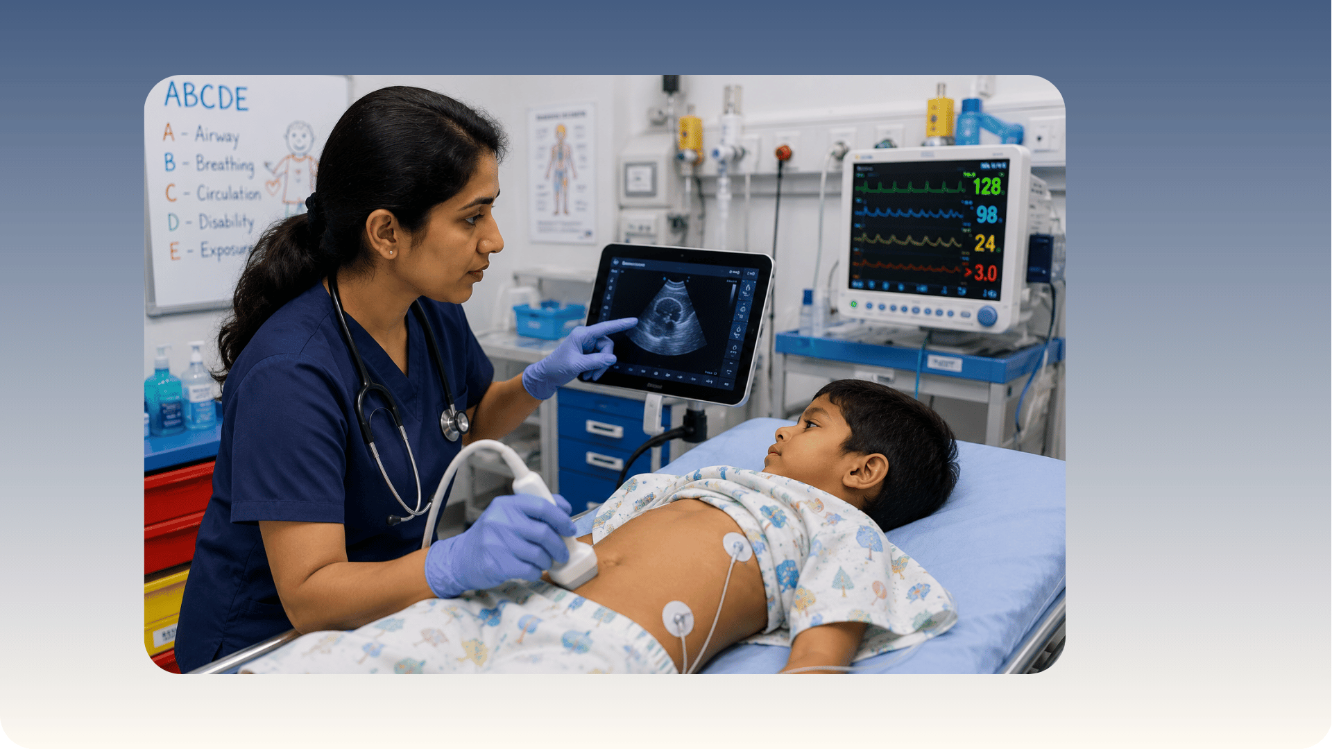
When a child arrives in the emergency department with difficulty breathing, abdominal pain, or a high fever and altered behaviour, the clinical question is always the same: what is happening inside this child, and how quickly can I know? For most of my career, answering that question meant waiting for a radiology report, a delay that in critical situations is time that cannot be afforded. Over the last several years, I have been using Point-of-Care Ultrasonography (POCUS) at the bedside to provide immediate answers in exactly these situations. As the doctor who introduced POCUS to paediatric practice in Eastern Uttar Pradesh, I want to explain what this technology actually does, how it is used in clinical care, and why it matters for children and families in Varanasi.
What is POCUS and how is it different from conventional ultrasound?
Most families are familiar with ultrasound from pregnancy scans or abdominal imaging. Conventional ultrasound is performed by a radiology department, involves scheduling and waiting, and produces a report that the treating doctor then receives and interprets. Point-of-Care Ultrasound is different in a fundamental way: it is performed by the treating clinician, at the bedside, in real time, as part of the clinical examination. The scan and the clinical decision happen in the same moment, rather than separated by a referral, a waiting period, and a report.
This distinction matters most in emergency and critical care settings, where the question being asked is not a comprehensive survey of an organ but a focused answer to a specific clinical question. Is there fluid around the heart? Are the lungs consolidating? Is this child's heart pumping adequately? POCUS answers these questions in minutes, without moving a critically ill child to a radiology suite, without radiation exposure, and without the delay that makes the difference in a deteriorating patient.
Why POCUS is particularly important in paediatric care
Children are not small adults. Their physiology is different, their ability to communicate symptoms is limited especially in infancy, and their conditions can progress more rapidly than in adults. A baby with sepsis may not show the classical signs of infection until the condition is already advanced. A toddler with respiratory distress cannot describe whether the difficulty is coming from the airway, the lungs, or the heart. Paediatric POCUS provides a window into these systems that physical examination alone cannot offer, and it does so without the radiation burden of X-ray or CT scanning, which is a genuine concern in children who may require multiple investigations over the course of a complex illness.
The lungs of a healthy child, the fluid status of a dehydrated infant, the cardiac function of a newborn with congenital disease, the presence of intussusception in a baby with unexplained colicky pain, all of these can be assessed rapidly and directly at the bedside with POCUS. In a setting like Varanasi, where families travel from across Eastern Uttar Pradesh and where access to specialist radiology is limited in the hours when children most commonly deteriorate, the ability to perform this assessment immediately without referral changes what is possible for the child in front of me.
Clinical applications: where POCUS makes the most difference
Lung and respiratory assessment
Respiratory conditions are among the most common reasons children require emergency assessment. A child who is breathing rapidly and using accessory muscles of breathing needs an immediate answer about the cause. Lung ultrasound in children can identify pneumonia, pleural effusion (fluid around the lung), pneumothorax (collapsed lung), and bronchiolitis patterns with accuracy that, for many of these conditions, rivals or exceeds chest X-ray.
In practice, this means that when a child arrives with respiratory distress, I can perform a lung scan within minutes of the examination and have a working answer before any other investigation has been ordered. A consolidation pattern in a child with fever and cough confirms pneumonia. A pattern of multiple B-lines, which are characteristic artefacts that appear when the lung surface is waterlogged, suggests bronchiolitis or fluid overload. A bright hyperechoic line without lung sliding on one side suggests pneumothorax. Each of these findings directs immediate management without waiting for an X-ray result.
Cardiac assessment
Cardiac causes of deterioration in children are often the most difficult to identify clinically and the most time-sensitive to treat. A newborn with congenital heart disease, a child with myocarditis, or a patient in septic shock with compromised cardiac output all require immediate information about how the heart is functioning. Bedside cardiac ultrasound in the paediatric emergency setting allows direct assessment of cardiac contractility, the presence of pericardial effusion (fluid around the heart), and chamber size, all in real time.
For families who have been told their child is in shock or has a cardiac concern, the ability to show them in real time what is happening with their child's heart, to explain what the scan is showing and what it means for the management, is part of the care that POCUS enables. It is not simply a diagnostic tool. It is a communication tool, and in a frightening situation that matters.
Assessment of dehydration and fluid status
Dehydration is one of the most common conditions requiring hospital admission in children, and its assessment has historically relied on clinical signs that are often unreliable, particularly in moderate dehydration. POCUS for dehydration in children allows direct measurement of the inferior vena cava, the major vein returning blood to the heart, which collapses with inspiration when a patient is significantly volume-depleted. This measurement provides an objective, reproducible assessment of fluid status that guides decisions about intravenous fluid administration with far greater precision than clinical signs alone.
In a child who is vomiting and unable to tolerate oral fluids, knowing objectively whether the circulation is compromised determines whether that child needs immediate intravenous access and fluid resuscitation or can be managed with careful oral rehydration therapy. This is a decision that POCUS helps me make with confidence at the bedside rather than on clinical estimation.
Abdominal assessment
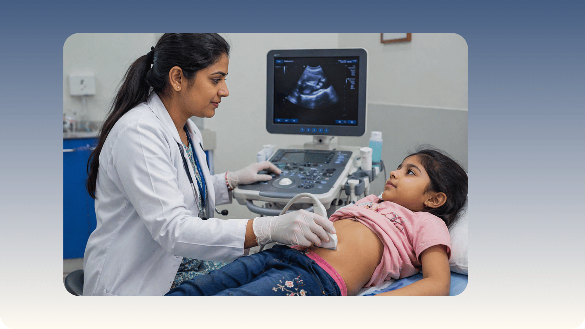
Abdominal pain in children is a common presentation with a broad differential diagnosis. In infants, intussusception, where one segment of bowel telescopes into another, is a surgical emergency that can look deceptively similar to a viral gastroenteritis in its early stages. Abdominal POCUS in paediatrics can identify the characteristic target sign of intussusception in minutes, allowing immediate referral for therapeutic enema or surgical management before the child deteriorates. Free fluid in the abdomen, appendiceal changes, and bowel wall thickening are also assessable with POCUS in appropriate cases.
In a setting where a radiology ultrasound may not be available for several hours, particularly at night and at weekends, POCUS provides a safety net for the recognition of surgical abdominal emergencies in children. I have used it on numerous occasions to identify findings that required immediate escalation of care, findings that would have remained undetected during a wait for formal imaging.
Procedural guidance
POCUS is also used to guide procedures, making them safer and more accurate. Ultrasound-guided procedures in children include central venous access, which is more safely performed when the target vein is visualised directly rather than located by anatomical landmarks alone; lumbar puncture, where ultrasound can identify the optimal intervertebral space particularly in a child with difficult anatomy; and thoracocentesis, the drainage of pleural fluid, where ultrasound locates the fluid pocket and guides the drainage needle away from lung tissue.
For parents, knowing that a procedure on their child is being performed under direct ultrasound visualisation rather than on anatomical estimation is a meaningful reassurance. It reduces the number of attempts required, reduces the risk of inadvertent injury to adjacent structures, and is associated with lower complication rates across all of these procedures.
POCUS in the neonatal setting
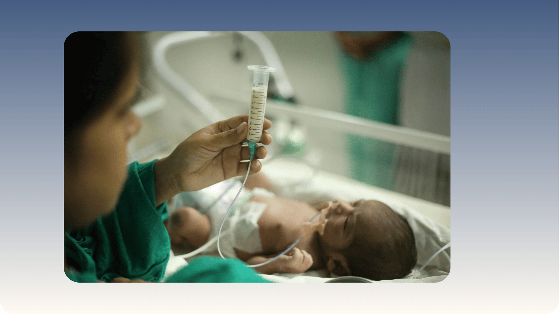
Newborns present particular diagnostic challenges because physical examination findings are subtle and symptoms are non-specific. A sick neonate may present with nothing more than poor feeding, temperature instability, or a change in colour. Neonatal POCUS allows rapid assessment of lung aeration in a baby with respiratory distress, cardiac function in a newborn with suspected congenital heart disease or persistent pulmonary hypertension, and cranial findings in a baby at risk of intraventricular haemorrhage.
In the neonatal period, the cranial fontanelle, the soft spot on the top of the skull that remains open in the early weeks of life, provides an acoustic window that allows ultrasound assessment of the brain without the need for CT scanning. This is particularly relevant in preterm infants who are at risk of intraventricular haemorrhage, a serious complication of prematurity that can be identified and monitored with serial cranial ultrasound at the bedside.
POCUS in resource-limited settings: why it matters for Eastern Uttar Pradesh
The impact of POCUS is greatest in settings where access to specialist radiology is limited, where families have travelled significant distances to reach care, and where delays in diagnosis carry the highest clinical cost. In the context of paediatric care in Varanasi and Eastern Uttar Pradesh, these conditions apply directly. A child who has been brought from a district three hours away cannot have their care delayed by a further wait for radiology. A sick infant in the early hours of the morning cannot be transferred to a radiology suite.
POCUS bridges this gap not by replacing specialist radiology but by providing the immediate focused answers that guide management in the time before specialist imaging is available, and by identifying conditions that require urgent escalation. In this way, it extends the reach of advanced diagnostic capability to the bedside of every patient, at any hour, without additional radiation and without a wait. This is the reason I invested in learning and introducing POCUS, and the reason I believe it should be part of the standard toolkit of every paediatric clinician working in emergency and critical care settings in India.
Training, competence, and the limits of POCUS
It is important to be clear about what POCUS is and what it is not. POCUS is a focused clinical tool, designed to answer specific questions at the bedside. It does not replace comprehensive formal ultrasonography performed by a trained radiologist, which remains the appropriate investigation for detailed organ assessment, characterisation of complex pathology, or when a comprehensive survey of an abdominal or thoracic structure is required. The value of POCUS lies in its speed, its portability, and its integration with the clinical examination, not in replacing the depth of assessment that formal radiology provides.
Competent use of POCUS requires specific training. I trained in POCUS through structured courses and have applied it in clinical practice for several years. The skills involved are not self-taught from general ultrasound exposure. They require supervised practice, assessment of image acquisition, and an understanding of the artefacts and pitfalls that can lead to misinterpretation. POCUS training for paediatricians is increasingly available in India through national training programmes, and its inclusion in paediatric emergency and critical care curricula is growing. As the clinician who introduced this practice in Eastern Uttar Pradesh, I have also been involved in sharing knowledge and supporting other clinicians who wish to develop these skills.
What POCUS means for families
For a parent watching their child in distress, waiting for answers is one of the most difficult parts of the experience. Bedside ultrasound for children means that in many clinical situations, that wait is significantly shorter. It means that a sick child does not have to be transported to another department, disconnected from monitoring, or delayed while a report is written. It means that the doctor examining your child can show you in real time what they are looking at and explain what they are seeing.
It also means that certain investigations that would previously have required referral, waiting, and in some cases overnight admission pending imaging, can now be performed and answered at the first point of contact. This has a direct impact on the efficiency of care, on the number of investigations a child requires, and on the length of hospital stay in many cases. It does not eliminate the need for formal radiology in complex cases, and I always refer for comprehensive imaging when the clinical question requires it. But for the focused questions that arise most frequently in paediatric emergency and critical care, POCUS has become as central to my examination as the stethoscope.
Questions parents ask me most often about POCUS
Is ultrasound safe for my child?
Yes. Ultrasound uses sound waves rather than radiation. There is no ionising radiation involved in ultrasound safety in children and no cumulative dose concern as there is with X-ray or CT scanning. Ultrasound has been used safely in clinical medicine for decades, including extensively in neonates and infants. Repeated POCUS assessments in a critically ill child carry no radiation burden, which is one of its most important advantages over other imaging modalities in paediatric care.
Will POCUS tell you everything you need to know?
POCUS answers specific, focused questions. It tells me whether there is fluid around the heart, whether the lungs are aerating normally, whether the bowel shows a target sign. It does not provide the comprehensive assessment of a formal radiology scan, and I use it as a complement to, not a replacement for, formal imaging when the clinical question requires it. The judgment about when POCUS is sufficient and when formal imaging is needed is part of the clinical skill involved in using this tool.
Does every paediatrician use POCUS?
POCUS is an emerging skill in paediatric practice in India and requires specific training. Not all paediatricians are trained in it. As the pioneer of POCUS in paediatric practice in Eastern Uttar Pradesh, I am one of a small number of clinicians in this region who perform this assessment routinely. This is part of what I believe makes the diagnostic approach at Vatsalya Children's Hospital different in the care we are able to offer to critically ill and acutely unwell children.
Can POCUS be used in newborns?
Yes, and it is particularly valuable in newborns. The open fontanelle provides a cranial window for brain assessment, and the small chest and abdomen of a POCUS in neonates makes cardiac, lung, and abdominal assessment highly feasible with appropriate equipment and technique. In premature and sick newborns where formal imaging poses challenges of transportation and radiation, POCUS at the bedside provides immediate information that guides critical management decisions.
If your child has been admitted with a serious illness or you would like a consultation with a paediatrician using POCUS as part of clinical assessment, please do not hesitate to contact us. To book a consultation with Dr Chhaya Pathak at Vatsalya Children's Hospital, Mahavir Road, Orderly Bazar, Varanasi, call +91 9838585111.
Written by Dr Chhaya Pathak, MBBS, MD (Pediatrics), Senior Consultant Paediatrician and Co-Founder, Vatsalya Children's Hospital, Mahavir Road, Orderly Bazar, Varanasi, Uttar Pradesh 221002. Pioneer of Paediatric Point-of-Care Ultrasonography (POCUS) in Eastern Uttar Pradesh.
Related reading
Pediatric Critical Care: Early Signs, Emergency Management, and Life-Saving Treatments for Children

Add a Comment