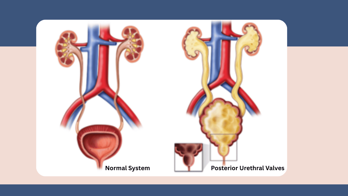
In my complete guide to neonatal surgery, I described posterior urethral valves as one of the urological emergencies that can present in the newborn period and require prompt specialist surgical assessment. Posterior urethral valves is also an area of my own research: during my MCh Paediatric Surgery training at the Institute of Medical Sciences, Banaras Hindu University, I studied aldosterone and renin levels as prognostic biomarkers in PUV patients, contributing to the understanding of how this condition affects the kidney's hormonal regulation. This personal and research connection to the condition means that when a family comes to Vatsalya Children Hospital, Varanasi with a son who has been diagnosed with PUV, either prenatally on an antenatal scan or in the newborn period, I understand not just the surgical management but the deeper biology of what the obstruction does to the kidneys over time, and what that means for the long-term care of these boys.
What Are Posterior Urethral Valves?
The urethra is the tube through which urine passes from the bladder to the outside of the body. In boys, this tube passes through the prostate gland before exiting at the tip of the penis. Posterior urethral valves are abnormal folds of tissue, present from birth, that form inside the urethra at the level of the prostate and obstruct the normal flow of urine out of the bladder. The obstruction is not complete in most cases, but it is sufficient to cause the bladder to work much harder than normal to push urine past the valves, producing a thickened, overactive bladder, and causing back-pressure on the kidneys through the ureters during a critical period of fetal renal development. PUV affects only males because only males have a posterior urethra in this anatomical location. PUV is the most common cause of severe obstructive uropathy in male newborns and one of the most significant causes of chronic kidney disease in childhood , not because the valves themselves are so destructive, but because the back-pressure they create during fetal life damages kidneys that are still developing.
Diagnosis: Antenatal Scan to Neonatal Assessment
The majority of significant PUV cases are now suspected before birth. A routine antenatal ultrasound scan showing a dilated bladder that does not empty fully, bilateral hydronephrosis (dilated kidneys), dilated ureters, and reduced amniotic fluid (oligohydramnios) in a male fetus raises the strong possibility of PUV. This is one of the reasons antenatal scanning matters so much , a PUV diagnosis made before birth allows the neonatal team to be prepared at delivery, investigations to begin immediately, and treatment to be initiated before the kidneys sustain additional postnatal damage. In a newborn with suspected PUV, a urethral catheter is placed as soon as possible to drain the bladder and relieve the obstruction acutely. A micturating cystourethrogram (MCUG), in which contrast dye is instilled into the bladder and X-ray images are taken during voiding, demonstrates the characteristic appearance of the dilated posterior urethra and confirms the diagnosis. Renal ultrasound and blood tests for creatinine and electrolytes assess the degree of existing kidney damage. The creatinine nadir, the lowest creatinine level reached in the weeks after birth as the maternal creatinine clears from the baby's blood, is one of the most reliable early indicators of the underlying kidney function.
Treatment: Valve Ablation and What Comes After
The definitive treatment for PUV is endoscopic valve ablation: a small cystoscope is passed through the urethra, and the obstructing valve tissue is divided using a fine diathermy hook or cold knife, removing the obstruction without any external incision. The procedure takes approximately 20 to 30 minutes under general anaesthesia and the catheter is removed within one to two days. In very small or premature newborns whose urethra is too narrow for the cystoscope, a temporary vesicostomy, a small opening from the bladder to the skin of the lower abdomen, may be performed first to divert urine and protect the kidneys until the child is large enough for endoscopic ablation. After ablation, the bladder and upper tracts are monitored with serial ultrasound scans to assess how the dilation resolves, and kidney function is monitored regularly with blood tests.
The surgical removal of the obstruction does not reverse damage that has already occurred to the kidneys or the bladder during fetal development. Many boys with PUV have some degree of bladder dysfunction even after successful valve ablation, because the bladder has spent months working against a high-resistance obstruction and may have developed abnormal patterns of storage and emptying that persist. Some boys have significant residual kidney impairment. Long-term follow-up with a paediatric urologist and nephrologist, bladder function assessment, and in some cases bladder training, anticholinergic medication, or intermittent catheterisation, are part of the ongoing care plan for boys with PUV. My research into aldosterone and renin as biomarkers in PUV contributes to the understanding of how the kidney's hormonal systems adapt to obstruction, and at Vatsalya Children Hospital, Varanasi, every boy treated for PUV is enrolled in a structured long-term follow-up programme, because the outcomes in this condition are determined as much by the quality of ongoing surveillance as by the quality of the initial surgery.
To book a consultation with Dr. Greeshma Suresh at Vatsalya Children Hospital, Mahavir Road, Orderly Bazar, Varanasi, Uttar Pradesh 221002, call +91 9838585111 or visit linqmd.com/doctor/greeshma-suresh
Written by Dr. Greeshma Suresh, MBBS, MS (General Surgery), MCh (Paediatric Surgery, Institute of Medical Sciences, Banaras Hindu University), Paediatric Surgeon, Vatsalya Children Hospital, Mahavir Road, Orderly Bazar, Varanasi, Uttar Pradesh 221002. 6+ years. 1,500+ surgical cases. Phone: +91 9838585111.
Related reading
Neonatal Surgery: A Complete Guide for Parents Facing a Newborn Surgical Diagnosis
Hirschsprung Disease in Newborns: What It Is, How It Is Diagnosed, and What Surgery Achieves
Congenital Anomalies in Newborns: Early Diagnosis and Pediatric Surgical Treatment Options


Add a Comment