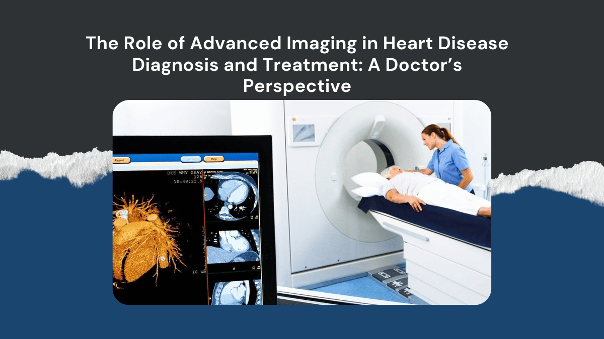
In my years of practice, I’ve come to realize that the difference between good and great patient care often lies in how precisely we can see the heart’s inner workings. Gone are the days when a simple angiogram was enough to tell us what we needed to know. Today, cutting-edge imaging technologies have transformed our ability to diagnose and treat heart disease with remarkable accuracy. Each detailed image becomes a story—an insight that guides us to the best possible path for each individual patient.
Transforming Diagnosis: Seeing Beyond the Surface
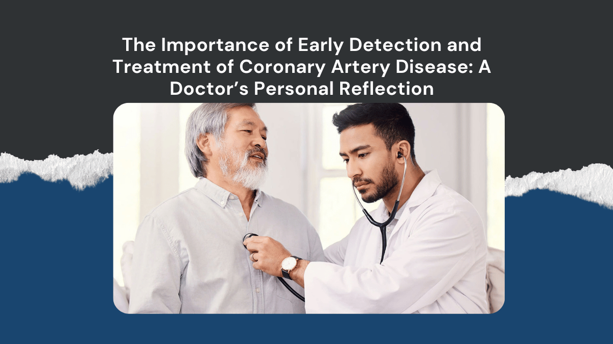
One patient, Mr. Kumar, a 58-year-old man with recurrent chest discomfort, came to me after multiple tests failed to provide a clear picture of his condition. Traditional angiograms showed some blockages, but they didn’t fully explain his symptoms. We then used advanced imaging techniques—specifically, cardiac MRI combined with 3D echocardiography. These images revealed subtle scarring in his heart muscle and hidden areas of stress that standard tests missed. That detailed insight allowed us to personalize his treatment plan, focusing on both medication and lifestyle changes that targeted the specific areas affected.
Why Advanced Imaging Matters
What makes advanced imaging so vital is its ability to visualize the heart beyond the limitations of conventional techniques. Cardiac MRI, for example, offers detailed tissue characterization, enabling us to identify early signs of myocarditis, cardiomyopathies, or scar tissue. 3D echocardiography, on the other hand, provides a more accurate assessment of valve function and chamber size—crucial details for complex cases. Cardiac CT scans give us a non-invasive window into coronary arteries, helping us detect plaques before they cause a heart attack.
For my patients, these tools mean earlier diagnosis, better risk assessment, and more effective, targeted treatments. It’s no longer just about managing symptoms; it’s about understanding the root cause with clarity and precision.
Patient Stories: Seeing is Understanding
Take Ms. Priya, a young woman with a family history of early heart disease. Standard tests showed her heart was fine, but I felt something was missing. We opted for a cardiac MRI, which revealed early signs of a different, less common condition—fibrotic tissue in her myocardium. That insight changed her management approach entirely, helping her avoid a potential future crisis and empowering her with knowledge and proactive care.
Similarly, in emergency situations, advanced imaging helps us act swiftly. I remember a patient with a suspected complex coronary blockage. A CT coronary angiogram revealed a critical lesion in a difficult-to-reach artery. This detailed imagery guided our intervention, minimizing risks and maximizing success.
Looking to the Future
As technology advances, I believe imaging will only become more integrated into every step of cardiac care. The future holds the promise of even more personalized diagnoses and treatments—perhaps combining imaging data with AI algorithms to predict heart events before they happen.
For patients, this means smarter, safer, and more effective care. For me, it’s a source of immense satisfaction to see the real-world impact—seeing patients regain their health, confidence, and even their lives, thanks to a clearer picture of their hearts.
Conclusion
Advanced cardiac imaging is not just a technological breakthrough; it’s a patient-centric approach that enhances our ability to diagnose accurately and treat effectively. It’s about seeing the unseen, understanding the unknown, and helping my patients lead healthier, happier lives.

Add a Comment