Skull base surgery is one of the most technically demanding disciplines in neurosurgery. It involves operating on tumours, vascular abnormalities, and other lesions located at the base of the brain, a region that houses critical structures including the brainstem, cranial nerves, major blood vessels, and the pituitary gland. As a Consultant Neurosurgeon specialising in skull base and vascular neurosurgery at Royal Preston Hospital, I have dedicated my career to this field, completing four international fellowships across the United Kingdom, United States, and Australia. This guide is for patients and families who want to understand what skull base surgery involves, what conditions it treats, and what to expect throughout the journey.
What is the skull base and why is it surgically challenging?
The skull base is the floor of the cranial cavity, the bony platform on which the brain rests. It is not a flat surface. It is a complex, three-dimensional structure with openings through which the cranial nerves and major blood vessels pass between the brain and the rest of the body. Conditions that develop in or around this region, tumours, vascular malformations, infections, or structural abnormalities, are among the most challenging in all of medicine to treat surgically.
The difficulty arises from anatomy. The skull base sits immediately adjacent to structures that control vision, hearing, facial sensation, swallowing, balance, and the fundamental functions of breathing and consciousness. Operating in this region requires a detailed understanding of three-dimensional surgical anatomy, advanced microsurgical technique, and the ability to navigate around structures that have no margin for error.
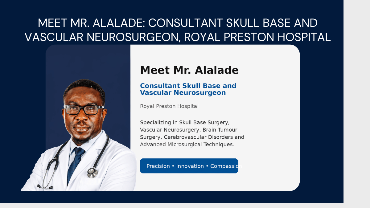
This is why skull base neurosurgery is a subspecialty within a subspecialty. Not all neurosurgeons operate at the skull base, and those who do typically complete dedicated fellowship training beyond standard neurosurgical residency. Mr. Alalade's own training included fellowships at The Walton Centre in Liverpool, Princess Alexandra Hospital in Brisbane, Weill Cornell Medical College in New York, and the National Hospital for Neurology and Neurosurgery in London.
What conditions are treated with skull base surgery?
Skull base surgery encompasses a wide range of conditions. The common thread is their location, at or near the base of the brain, and the complexity of the surgical approach required to address them safely.
Acoustic neuroma (vestibular schwannoma)
Acoustic neuroma surgery addresses a benign tumour that grows on the vestibulocochlear nerve, the nerve responsible for hearing and balance. Despite being benign, these tumours can cause progressive hearing loss, tinnitus, facial numbness, and balance problems as they grow. They can also compress the brainstem if they reach a significant size. Treatment options include observation with regular MRI monitoring, stereotactic radiosurgery, and microsurgical removal. The choice depends on tumour size, rate of growth, the patient's hearing status, and their overall clinical picture. In surgical cases, the approach, retrosigmoid, translabyrinthine, or middle fossa, is selected based on the individual's anatomy and the goal of treatment.
Meningioma of the skull base
Meningiomas are the most common primary brain tumour. Those arising from the skull base are among the most surgically challenging because of their proximity to critical neurovascular structures. Skull base meningiomas can compress cranial nerves, major arteries, the brainstem, or the cavernous sinus. Many grow slowly and are found incidentally on imaging. Surgical decisions depend on growth rate, symptoms, the degree of involvement of surrounding structures, and the patient's age and overall health. Advances in microsurgical technique and neuronavigation have significantly improved the safety of skull base meningioma surgery over the past two decades.
Pituitary tumours
The pituitary gland sits in the sella turcica, a bony depression at the centre of the skull base. Pituitary tumour surgery addresses the most common skull base pathology managed operatively. These tumours may be non-functioning, causing symptoms only through compression of surrounding structures, or functioning, producing excess hormones that cause conditions including acromegaly, Cushing's disease, and hyperprolactinaemia. The standard surgical approach is endoscopic transsphenoidal surgery, passing instruments through the nostril and nasal cavity to reach the pituitary without any external incision. Mr. Alalade performs this using advanced endoscopic techniques developed in part through his research fellowship at Weill Cornell Medical College in New York.
Chordoma and chondrosarcoma
Chordomas are rare, slow-growing tumours that arise from remnants of the notochord, the embryonic structure from which the spine develops. They most commonly occur at the skull base or in the sacrum. Despite being locally aggressive, they rarely metastasise. Complete surgical removal is the goal of treatment but is technically demanding given the location at the clivus and the proximity to the brainstem and major vessels. Chondrosarcomas are similarly challenging. Both conditions require a subspecialist with specific experience in clival surgery.
Trigeminal neuralgia
Trigeminal neuralgia is characterised by episodes of severe, electric shock-like facial pain along the distribution of the trigeminal nerve. In many cases, it is caused by a blood vessel compressing the trigeminal nerve root at the skull base. Microvascular decompression, a surgical procedure that separates the offending vessel from the nerve, is the most effective treatment for eligible patients. The procedure requires access to the posterior fossa through a small craniotomy and produces long-term pain relief in a significant proportion of patients. Endoscopic skull base surgery techniques have expanded the options available for many of these conditions, reducing surgical trauma and recovery time.
Cerebral aneurysm and neurovascular conditions
Cerebral aneurysm treatment forms a significant part of Mr. Alalade's vascular neurosurgery practice alongside skull base surgery. Arteriovenous malformations, abnormal tangles of blood vessels that can bleed into the brain, are also managed within this subspecialty. Aneurysms at the skull base, including those of the internal carotid artery and the basilar artery, are among the most complex to treat surgically and require both open microsurgical clipping and endovascular coiling options to be available and expertly performed.
How is skull base surgery approached, what techniques are used?
Skull base surgery is not a single operation. It is a collection of approaches tailored to the specific location of the pathology, the goals of surgery, and the individual patient's anatomy.
Microsurgical craniotomy approaches
A skull base craniotomy involves removing a section of the skull to access the brain. The approach is defined by the location of the lesion. The retrosigmoid approach accesses the posterior fossa and cerebellopontine angle, the region where acoustic neuromas and posterior fossa meningiomas are found. The pterional and orbitozygomatic approaches access the anterior and middle skull base. The transpetrosal approach accesses the petroclival region for lesions involving the clivus and petrous bone. Each approach is designed to provide the optimal viewing angle and surgical corridor while minimising brain retraction and preserving critical structures.
Endoscopic skull base surgery
Endoscopic skull base surgery uses a thin, rigid endoscope passed through the nostril to access the skull base without external incision. Originally developed for pituitary surgery, it has expanded significantly to include approaches to the anterior skull base, the clivus, the cavernous sinus, and the craniovertebral junction. Mr. Alalade's research fellowship in minimally invasive endoscopic brain and skull base surgery at Weill Cornell Medical College in New York was specifically focused on advancing these techniques. Endoscopic surgery reduces surgical trauma, eliminates external scarring, and shortens recovery time compared to open craniotomy for appropriate lesions.
Combined approaches
For complex skull base pathology, large tumours that extend across multiple anatomical compartments, or lesions involving both the intradural and extradural spaces, combined approaches that use both craniotomy and endoscopic access may be required. These are the most technically demanding cases in neurosurgery and are best managed in specialist skull base centres with a dedicated multidisciplinary team.
What does a multidisciplinary skull base team involve?
Complex skull base conditions are rarely managed by a neurosurgeon alone. At Royal Preston Hospital, skull base cases are reviewed by a multidisciplinary brain tumour team that brings together neurosurgery, neuroradiology, neuropathology, ear nose and throat surgery, ophthalmology, endocrinology, radiation oncology, and neuro-oncology. This team approach ensures that every patient's case is reviewed by the relevant specialists before a management plan is recommended.
The team also includes clinical nurse specialists and allied health professionals who support patients through the investigation, treatment, and recovery process. For patients with pituitary tumours, close collaboration with endocrinology is essential both before and after surgery. For patients with acoustic neuromas, a joint assessment with neurotology helps determine the optimal approach to preserving hearing where possible.
What are the risks of skull base surgery?
Skull base surgery carries risks that are specific to the procedures involved and the individual patient's anatomy. Mr. Alalade discusses these in detail at the pre-operative consultation, and does not recommend surgery without ensuring that the patient and their family have a clear understanding of what the procedure involves and what the realistic risks are.
Cranial nerve injury
The skull base contains twelve pairs of cranial nerves controlling vision, eye movement, facial sensation, facial movement, hearing, balance, taste, swallowing, and voice. Understanding cranial nerve surgery risks is central to informed consent for any skull base procedure. Facial nerve monitoring, auditory brainstem response monitoring, and other intraoperative neurophysiological monitoring tools are used to protect these structures during surgery. Temporary cranial nerve deficits, weakness, numbness, or altered function, are common after skull base surgery and often improve over weeks to months as the nerve recovers.
CSF leak
Cerebrospinal fluid leak, where the fluid surrounding the brain escapes through the surgical site, is a recognised complication particularly in endoscopic skull base surgery. It is managed with careful surgical closure using tissue grafts, fat, and fibrin sealant, and in most cases resolves with conservative management or a minor surgical repair.
Vascular injury
The skull base is traversed by major intracranial arteries and venous sinuses. Injury to these structures is rare but serious. Meticulous surgical planning using preoperative imaging and angiography, combined with intraoperative neuronavigation, minimises this risk.
What does recovery from skull base surgery look like?
Hospital stay and early recovery
Recovery varies considerably depending on the specific procedure performed, the size and location of the pathology, and the patient's pre-operative neurological status. For open craniotomy procedures, skull base surgery recovery typically involves a hospital stay of five to ten days. Patients are monitored in a high-dependency or intensive care setting immediately after surgery. Headache, fatigue, and mild neurological symptoms in the early post-operative period are common and usually improve as swelling settles over the first two to four weeks.
Rehabilitation
Patients with cranial nerve deficits after skull base surgery, facial weakness, hearing change, swallowing difficulty, or balance problems, are referred for appropriate brain tumour rehabilitation. This may include facial physiotherapy, speech and language therapy, vestibular rehabilitation, or audiological assessment and hearing aid fitting. Rehabilitation significantly improves long-term functional outcomes and is an integral part of the care Mr. Alalade provides rather than an optional add-on.
Long-term follow-up
Skull base conditions, including meningiomas, acoustic neuromas, pituitary tumours, and chordomas, require skull base tumour follow-up with regular MRI to monitor for recurrence or regrowth. The follow-up schedule depends on the condition, the extent of surgical resection, and whether adjuvant treatment such as stereotactic radiosurgery or radiotherapy was used. Mr. Alalade discusses the long-term follow-up plan with every patient before they leave hospital.
When should you seek a skull base neurosurgery opinion?
A skull base neurosurgeon consultation is appropriate when imaging has identified a lesion at or near the skull base, including the posterior fossa, pituitary region, cerebellopontine angle, clivus, or cavernous sinus, or when symptoms suggest cranial nerve involvement such as progressive hearing loss, tinnitus, facial numbness, double vision, swallowing difficulty, or facial pain.
A referral is also warranted when a diagnosis of acoustic neuroma, meningioma, pituitary tumour, chordoma, or cerebral aneurysm has been made and specialist advice on management is required, when a second opinion on a complex skull base case is being sought, or when previous skull base surgery has resulted in recurrence or residual tumour requiring further management.
An opinion from Mr. Alalade does not automatically lead to surgery. Many skull base conditions are managed conservatively with surveillance, and the consultation is as much about understanding the full picture and all available options as it is about planning an operation.
Questions patients ask most often about skull base surgery
Is skull base surgery always necessary for a tumour at the base of the brain?
No. Many skull base tumours, particularly small acoustic neuromas, incidentally discovered meningiomas, and non-functioning pituitary adenomas, can be safely managed with active surveillance using regular MRI monitoring. Surgery is recommended when there is evidence of growth, when symptoms are developing or worsening, or when the size or location of the tumour poses a risk to critical structures. The decision is always based on the full clinical picture, not the imaging findings alone.
What is the difference between microsurgery and radiosurgery for skull base tumours?
Microsurgery vs radiosurgery is one of the most common questions Mr. Alalade addresses in skull base consultations. Microsurgery involves physically removing the tumour through a surgical approach to the skull base. Radiosurgery, such as Gamma Knife or CyberKnife, delivers a precisely focused dose of radiation to the tumour, aiming to stop its growth rather than remove it physically. Both have established roles in skull base tumour management. The choice depends on tumour size, location, the patient's age and health, hearing status, and the goals of treatment. In many cases, a combination of surgery and radiosurgery is the optimal approach, particularly for large tumours or residual disease after incomplete resection.
How do I know if my surgeon has the experience needed for a complex skull base case?
When seeking a consultation for a complex skull base condition, it is reasonable to ask about your surgeon's specific experience with that condition, the number of cases they perform annually, and whether they operate within a dedicated multidisciplinary skull base team. Surgeons with dedicated fellowship training in skull base surgery from internationally recognised centres, and those who publish in the field, bring a depth of clinical knowledge that extends beyond standard neurosurgical training.
Can skull base tumours recur after surgery?
Yes, recurrence is possible, particularly for conditions such as meningioma, chordoma, and pituitary adenoma. The risk depends on the extent of surgical resection, the tumour grade, and the specific pathology. Complete surgical removal reduces recurrence risk but is not always achievable given the proximity of skull base tumours to critical structures. Long-term MRI surveillance and, in selected cases, adjuvant radiosurgery or radiotherapy form part of the comprehensive management plan.
Written by Mr. Andrew F. Alalade, MBBS, MRCS, FRCS (SN), FEBNS, Consultant Neurosurgeon (Skull Base and Vascular), Royal Preston Hospital, Lancashire, United Kingdom. Associate Editor, British Journal of Neurosurgery. Author of 100+ peer-reviewed publications.
Citations:
- Almefty KK, Pravdenkova S, Colli BO et al. Chordoma and chondrosarcoma: similar, but quite different, skull base tumors. Cancer, 2007.
- Komotar RJ, Starke RM, Raper DM et al. Endoscopic skull base surgery: a comprehensive comparison with open transcranial approaches. British Journal of Neurosurgery, 2012.
Related reading:
Acoustic Neuroma Treatment, Preston, Lancashire
Cerebral Aneurysm Treatment, Preston, Lancashire
Pituitary Tumour Surgery, Preston, Lancashire
Brain Tumour Surgery, Preston, Lancashire

Add a Comment