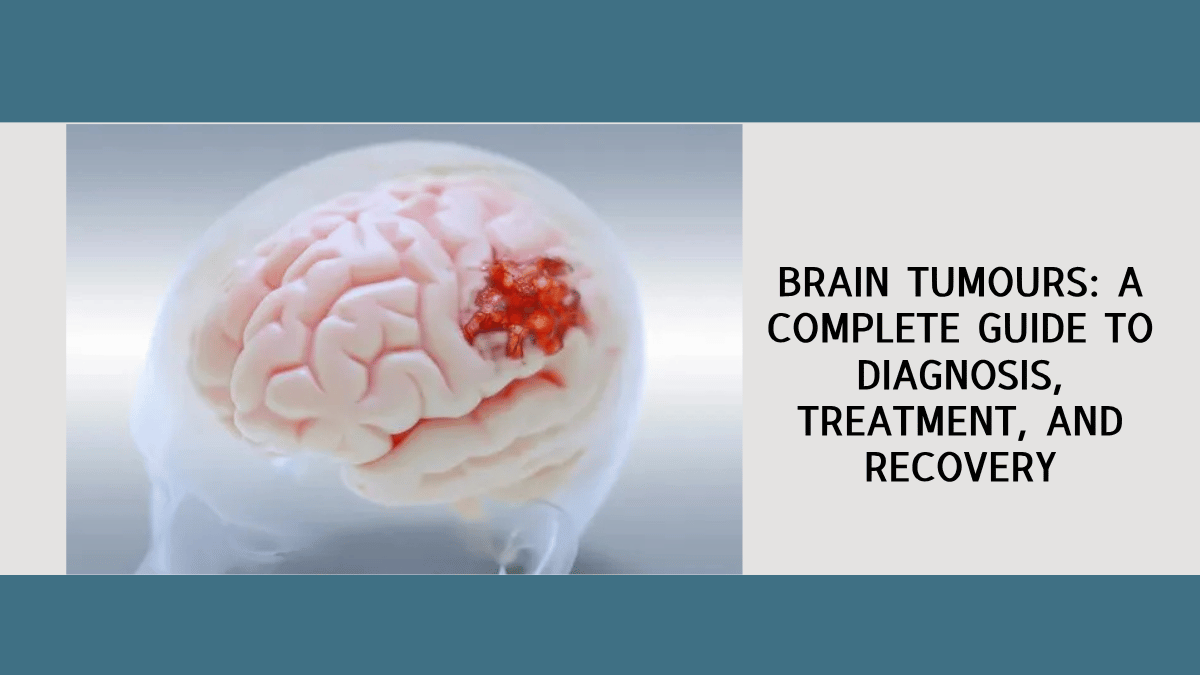
A brain tumour diagnosis is one of the most frightening things a person or a family can hear. In my experience as a neurosurgeon, the fear that follows a diagnosis is almost always worse than the reality of what treatment involves, and it is made worse by the fact that the information available online is often generic, frightening, and disconnected from the specific situation the patient is actually in. The range of outcomes in brain tumour surgery is genuinely wide. Some tumours are benign, slow-growing, and curable with a single well-executed operation. Others are more aggressive and require a multimodal approach involving surgery, radiation, and chemotherapy. And some can be managed conservatively with observation and monitoring, without any surgery at all. I am Dr. Prashanth Alle, Consultant Neurosurgeon at Apex Hospital and Gudage Hospital in Bidar, Karnataka, with DNB Neurosurgery from Sri Sathya Sai Institute of Higher Medical Sciences, Bengaluru, ECFMG certification from the United States, and 18,200 patients across my career. My training and publications span glioma, meningioma, pituitary adenoma, paediatric brain tumours, and craniovertebral junction anomalies, and this guide is written to give every patient in Bidar and across northern Karnataka the clear, specific information about brain tumours that they deserve before they walk into a neurosurgeon's consultation room.
What Is a Brain Tumour?
A brain tumour is an abnormal growth of cells within or adjacent to the brain. Tumours can arise from the brain tissue itself (primary brain tumours), from the membranes covering the brain (meningiomas), from the nerves entering the skull (schwannomas and neuromas), from the pituitary gland, or from cancer elsewhere in the body that has spread to the brain (metastatic or secondary brain tumours). The term brain tumour does not mean the same thing as brain cancer. Many brain tumours are benign, meaning they do not invade surrounding tissue or spread to other parts of the body, though they can still cause serious symptoms and require treatment by virtue of their location. Understanding this distinction is the first and most important step, because the specific type, grade, and location of a brain tumour determines everything about its treatment and prognosis.
Types of Brain Tumours: What the Diagnosis Actually Means
Gliomas
Gliomas arise from the glial cells of the brain, which are the supporting cells that surround and protect neurons. They range from low-grade gliomas (Grade 1 and 2), which grow slowly and may be managed with surgery alone in some cases, to high-grade glioblastomas (Grade 4), which are aggressive and require a combination of maximal safe surgical resection, radiotherapy, and chemotherapy. The grade of a glioma is determined by pathological examination of the tumour tissue after surgery, not by imaging alone. A glioma appearing on MRI may look very different from another glioma that carries the same radiological appearance but a completely different molecular profile, and it is the molecular markers, particularly IDH mutation status and MGMT methylation, that most powerfully influence the treatment strategy and the prognosis. Maximally safe resection of glioma, guided by neuronavigation and intraoperative imaging, is the standard first step in management and significantly influences both survival and quality of life.
Meningiomas
Meningiomas arise from the meninges, the three-layered membrane that covers the brain and spinal cord, and represent the most common primary intracranial tumour. They are classified by their location: convexity meningiomas on the surface of the brain, parasagittal and falcine meningiomas adjacent to the large venous sinus, skull base meningiomas involving the floor of the skull and critical neural structures, and posterior fossa meningiomas near the cerebellum and brainstem. The majority of meningiomas are benign (WHO Grade 1) and grow slowly, sometimes remaining asymptomatic for years. The decision to operate depends on the size, location, rate of growth on serial imaging, and the patient's symptoms and age. When surgery is indicated, the goal is complete resection, which in accessible locations produces a high cure rate. My training at Sathya Sai Institute included extensive exposure to all meningioma subtypes, including the complex skull base and posterior fossa locations where the surgical approach requires detailed anatomical knowledge and precise microsurgical technique.
Pituitary Adenomas
Pituitary adenomas are tumours of the pituitary gland, the small but critically important endocrine gland at the base of the brain that regulates growth hormone, thyroid function, adrenal function, and reproductive hormones. They may be functional, meaning they secrete hormones in excess and cause specific endocrine syndromes (Cushing's disease from ACTH excess, acromegaly from growth hormone excess, hyperprolactinaemia from prolactin excess), or non-functional, causing symptoms purely by mass effect including headache, visual field defects from pressure on the optic chiasm, and pituitary insufficiency from compression of normal gland tissue. My publication in World Neurosurgery in 2024, examining the optico-chiasmatic distortions produced by pituitary adenomas and the relationship between postoperative morphological reversal and visual recovery, reflects the depth of clinical and research interest I bring to the management of these tumours. Endoscopic transnasal surgery for pituitary macroadenoma allows the tumour to be removed through the nasal cavity without any external incision, with excellent visualisation of the tumour and the surrounding critical structures.
Posterior Fossa Tumours in Adults and Children
The posterior fossa is the compartment at the back and base of the skull, housing the cerebellum, the brainstem, and the fourth ventricle. Tumours in this location are particularly important in children, where medulloblastoma and ependymoma are the most common, and in adults where acoustic neuromas (vestibular schwannomas), meningiomas, and haemangioblastomas predominate. Symptoms from posterior fossa tumours include headache from raised intracranial pressure due to hydrocephalus, balance problems and unsteadiness, double vision, and difficulty swallowing. My publication in Childs Nervous System in 2024 on primary intracranial paediatric ganglioneuroblastoma reflects specific clinical experience with unusual and diagnostically challenging posterior fossa tumour presentations in children.
Cerebral Metastases
Metastatic brain tumours are actually the most common intracranial tumours, arising from cancers elsewhere in the body (lung, breast, melanoma, colorectal, and renal are the most frequent sources) that have spread to the brain through the bloodstream. Their management requires close collaboration with the oncology team and varies with the number and location of metastases, the status of the primary cancer, and the patient's overall performance status. Surgery may be appropriate for a single or dominant accessible metastasis, either alone or combined with stereotactic radiosurgery to the remaining lesions.
Symptoms of a Brain Tumour: What to Watch For
The symptoms of a brain tumour are determined largely by the location of the tumour and its rate of growth. A slowly growing meningioma may produce no symptoms for years, discovered incidentally on an MRI performed for another reason. A rapidly growing high-grade glioma may produce symptoms that progress over days to weeks. The most common symptoms that should prompt neurological evaluation include: new persistent headaches, particularly those that are worse in the morning or disturb sleep; seizures in an adult with no prior history of epilepsy; progressive weakness, numbness, or clumsiness on one side of the body; speech difficulty, whether in finding words or in articulating them clearly; vision changes, particularly in the visual field of one or both eyes; personality or behaviour change that is noticed by family before the patient acknowledges it; and difficulty with memory, concentration, or complex thinking. A new seizure in an adult is a brain tumour until proven otherwise, and requires immediate neurological evaluation.
How a Brain Tumour Is Diagnosed
The diagnosis of a brain tumour begins with clinical evaluation and is confirmed by neuroimaging, typically an MRI with gadolinium contrast enhancement. MRI provides the anatomical detail needed to characterise the tumour: its location, size, degree of surrounding oedema, relationship to critical structures such as motor cortex and speech areas, and the presence of hydrocephalus or herniation. CT scan is more rapidly available and remains the first imaging in an emergency presentation, particularly when haemorrhage into the tumour is suspected.
Advanced imaging including MR spectroscopy, functional MRI for mapping eloquent cortex, and diffusion tensor imaging for mapping white matter tracts adds further information that is critical for surgical planning in tumours adjacent to motor or speech areas. For most primary brain tumours, histopathological and molecular analysis of the tumour tissue obtained at surgery provides the definitive diagnosis, the tumour grade, and the molecular markers that guide adjuvant treatment decisions. The MRI is the beginning of the diagnostic process, not the end , the specific tissue diagnosis that surgery provides is what determines the full treatment plan.
Brain Tumour Surgery: What the Operation Involves
The prospect of brain surgery is frightening for most patients, and the questions I am asked most often in my pre-operative consultations reflect this: Will I be awake? Will it change who I am? Will I wake up with weakness? These are important questions that deserve specific, honest answers based on the individual patient's tumour location and characteristics, not generic reassurances.
Craniotomy for Brain Tumour Removal
A craniotomy involves making a temporary opening in the skull bone, accessing the brain through the opening with microsurgical instruments under high magnification, removing the tumour, and replacing the bone. The critical principle governing brain tumour surgery is maximal safe resection: removing as much of the tumour as safely possible while preserving the neurological function of the surrounding brain. This balance is achieved through the use of neuronavigation, which provides real-time MRI guidance during surgery to help the surgeon know exactly where within the brain the instruments are; intraoperative neurophysiological monitoring, which tracks motor and sensory signals from critical pathways during surgery to provide early warning if the dissection is approaching a functional area; and in selected tumours adjacent to speech areas, awake craniotomy, where the patient is awake during part of the procedure to allow real-time testing of language function. Neuronavigation and intraoperative monitoring have transformed brain tumour surgery from a procedure guided by anatomical landmarks alone to one guided by real-time physiological and imaging data.
Endoscopic and Minimally Invasive Approaches
For pituitary tumours and selected intraventricular and skull base lesions, endoscopic approaches through the nasal cavity or through small cranial openings allow tumours to be removed with minimal disruption to surrounding brain tissue. Endoscopic third ventriculostomy, a procedure I perform for hydrocephalus caused by tumour obstruction of CSF pathways, creates an alternative drainage route for CSF without placing an external shunt. These minimally invasive approaches, when appropriate for the clinical situation, reduce hospital stay and recovery time significantly compared with open craniotomy.
After Brain Tumour Surgery: Recovery and Adjuvant Treatment
Most patients who undergo brain tumour surgery spend several days in hospital after the procedure, initially in a monitored neurosurgical environment and then on the neurosurgical ward as they mobilise and begin recovery. The degree and rate of recovery depend on the tumour type, the extent of resection, the location, and the patient's baseline neurological status.
For malignant gliomas, surgery is followed by concurrent radiotherapy and chemotherapy (temozolomide), typically beginning four to six weeks after the operation once the wound has healed and the patient has recovered sufficient function. For benign tumours such as meningiomas and pituitary adenomas that have been completely excised, no further treatment may be required, with monitoring by serial MRI. For acoustic neuromas, the decision between surgery, stereotactic radiosurgery, and observation depends on the size of the tumour, the rate of growth, and the patient's hearing and facial nerve status. The treatment plan after brain tumour surgery is individualised based on the pathological findings, the extent of resection, and the patient's recovery and is made jointly by the neurosurgeon, oncologist, and radiation oncologist in a multidisciplinary team meeting.
Brain Tumour Surgery in Bidar: Access to Advanced Neurosurgery in Northern Karnataka
Bidar is the northernmost district of Karnataka, and patients from this region have historically had to travel to Hyderabad or Bengaluru for neurosurgical care. The presence of an advanced neurosurgical service in Bidar changes that equation significantly. At Apex Hospital and Gudage Hospital, I offer the full range of neurosurgical management for brain tumours: craniotomy for glioma, meningioma, and metastases; endoscopic transnasal resection for pituitary macroadenomas; posterior fossa surgery for cerebellar and brainstem tumours; and paediatric neurosurgical care including brain tumour resection in children. My DNB training at Sathya Sai Institute gave me exposure to the full complexity of neurosurgical cases in one of India's most respected neurosurgical programmes, and my six peer-reviewed publications reflect the academic rigour I continue to bring to clinical decision-making. If you or a family member in Bidar or the surrounding districts has been told that a brain tumour has been found on imaging, the first step is a neurosurgical consultation. That consultation, with a neurosurgeon trained in the full spectrum of intracranial tumour surgery, is available in Bidar at Apex Hospital and Gudage Hospital.
To book a neurosurgery consultation with Dr. Prashanth Alle at Apex Hospital, Bidar (Monday to Saturday, 11 AM to 2 PM, call +91 9036360396) or Gudage Hospital, Bidar (Monday to Saturday, 3 PM to 5 PM, call +91 84822 26841).
Written by Dr. Prashanth Alle, MBBS (M.S. Ramaiah Medical College, Bengaluru), ECFMG (USA), DNB Neurosurgery (Sri Sathya Sai Institute of Higher Medical Sciences, Bengaluru), Consultant Neurosurgeon, Apex Hospital, Bidar (Monday to Saturday, 11 AM to 2 PM) and Gudage Hospital, Bidar (Monday to Saturday, 3 PM to 5 PM), Karnataka. 18,200+ patients. Phone: +91 9632384477.
Related reading
World Brain Day: When Your Brain Sends a Warning, Listen Early
Head Injuries and Brain Bleeds When to Rush to a Neurosurgeon


Add a Comment