
In my complete guide to breast lumps, I described fibroadenoma as the most common benign breast lump in younger women, one that often feels smooth, firm, and mobile beneath the skin. One of the most frequent questions I receive after a woman has been told she has a fibroadenoma is whether she needs surgery. It is a reasonable question, and it does not have a single answer that applies to everyone. Whether a fibroadenoma should be removed depends on its size, how it is behaving over time, what the investigations show, and what the woman herself wants. Understanding the factors that guide that decision is the purpose of this blog.
What Is a Fibroadenoma?
A fibroadenoma is a benign tumour composed of glandular and connective breast tissue. It is not cancer, it does not become cancer in the large majority of cases, and it does not spread to other parts of the body. It typically presents as a well-defined, painless, movable lump that a woman discovers herself or that is found incidentally on imaging. Fibroadenomas are most common in women in their teens, twenties, and thirties, and are related to oestrogen sensitivity in breast tissue, which is why they often enlarge slightly during pregnancy and shrink after menopause. They can be single or multiple, and they can occur in one or both breasts. The diagnosis of a fibroadenoma should always be confirmed through appropriate investigation rather than clinical examination alone, because other breast lumps, including phyllodes tumours, which can resemble fibroadenomas clinically, require a different management approach.
How Is a Fibroadenoma Diagnosed?
The standard investigation pathway for a suspected fibroadenoma begins with clinical examination and ultrasound imaging. In younger women, ultrasound is typically the first-line imaging modality for breast lumps. A fibroadenoma has characteristic features on ultrasound: a well-defined, oval, hypoechoic lesion with smooth margins and uniform internal echoes. In women over 35, mammography is often added to the assessment. When imaging findings are equivocal or when the lump has concerning features, a core needle biopsy provides a tissue diagnosis, which is the most reliable way to confirm that a lump is a fibroadenoma and not something else. At my practice in Bengaluru, the triple assessment approach of clinical examination, imaging, and tissue sampling where indicated is the foundation of evaluating any breast lump.
When Can a Fibroadenoma Be Monitored Without Surgery?
Many fibroadenomas do not need to be removed. A small fibroadenoma that has been confirmed on imaging and biopsy to be a simple fibroadenoma, that is not growing, and that is not causing significant symptoms can often be managed with periodic clinical and ultrasound review rather than surgery. Over time, some fibroadenomas remain stable, some shrink spontaneously, and some slowly enlarge. Regular monitoring allows the clinical team to detect any change in behaviour early and adjust the management plan accordingly. This approach is appropriate for carefully selected patients who are comfortable with monitoring and who understand what changes should prompt them to return for review before their next scheduled appointment. Monitoring is not the same as ignoring a lump. It is a structured, active decision that requires clear follow-up arrangements and patient awareness of what to watch for.
When Is Surgical Removal the Right Decision?
Surgical removal of a fibroadenoma becomes the appropriate recommendation in several specific situations. When a fibroadenoma is enlarging on serial imaging, the growth itself is a reason to reconsider the monitoring approach. When a lump is large enough to be causing physical discomfort, distortion of breast shape, or significant psychological distress, these are legitimate indications for removal. When imaging or biopsy results are not entirely consistent with a simple fibroadenoma, tissue removal for complete histopathological assessment provides the definitive answer. When a woman has a strong personal preference for removal despite a genuinely benign diagnosis and a clear explanation of the monitoring alternative, her preference is a valid part of the decision. At BOSH and Motherhood Hospital in HRBR Layout, Bengaluru, I perform fibroadenoma excisions as planned surgical procedures with careful incision planning designed to minimise visible scarring, and the removed tissue is always sent for histopathological examination to confirm the diagnosis.
To book a consultation with Dr. Anupama Pujar K at BOSH HRBR Layout (Monday, Wednesday, Thursday 5:00 PM to 6:00 PM; Saturday 9:00 AM to 10:00 AM, call 080672 38855), Motherhood Hospital HRBR Layout (Tuesday, Friday 5:00 PM to 6:00 PM, call 080 6954 9278), or Sapthagiri Super Speciality Hospital, Hesarghatta Road (Tuesday, Friday 9:00 AM to 3:00 PM, call +91 9108475968), visit linqmd.com/doctor/anupama-pujar-k
Written by Dr. Anupama Pujar K, MBBS, MS General Surgery, FMAS, FIAGES, Dip MAS, ATLS Provider, General and Laparoscopic Surgeon, Bangalore Orthopaedic and Surgical Hospital (BOSH) HRBR Layout, Motherhood Hospital HRBR Layout, and Sapthagiri Super Speciality Hospital, Bengaluru. Phone: +91 80 6954 9278 / +91 80672 38855.
Related reading
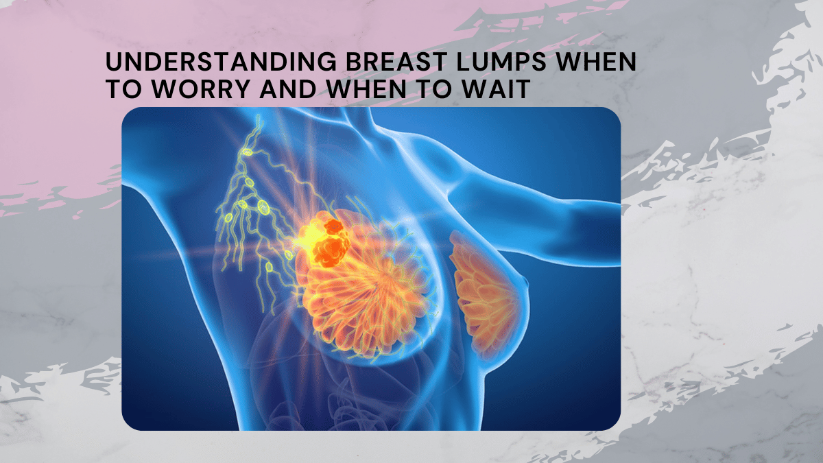
Understanding Breast Lumps When to Worry and When to Wait
The Importance of Early Detection in Gastrointestinal Disorders from Dr Anupama Pujar K

Add a Comment