Hydrocephalus and VP Shunt Surgery: What Patients and Families Need to Know About Anaesthesia and Care
When a family is told that their child or adult relative has hydrocephalus and requires a ventriculoperitoneal shunt, the questions that follow are almost always about the surgery itself: what happens in the operating theatre, what the risks are, and what recovery looks like. As a neuroanaesthesiologist at Sparsh Hospital, Hennur, Bengaluru, with 19 years of experience managing anaesthesia for neurosurgical procedures, I want to address the questions that families often bring to me about Hydrocephalus symptoms, the surgical options, and specifically what the anaesthesia team's role is in keeping a patient with raised intracranial pressure safe.
What is hydrocephalus and why does it require surgical treatment?
Hydrocephalus is an abnormal accumulation of cerebrospinal fluid (CSF) within the brain's ventricular system. CSF is produced continuously by the choroid plexus, circulates around the brain and spinal cord, and is absorbed into the venous system. When this circulation is obstructed, when absorption is impaired, or when production is excessive, fluid accumulates, the ventricles enlarge, and intracranial pressure rises. In infants whose skull sutures have not yet fused, this produces head enlargement. In older children and adults, where the skull is fixed, the pressure rise produces headache, nausea, vomiting, visual changes, cognitive deterioration, and in severe cases deterioration of consciousness. Without Hydrocephalus treatment, the condition is progressive and potentially life-threatening.
Hydrocephalus may be congenital, present from birth due to aqueductal stenosis, neural tube defects, or chromosomal anomalies, or acquired following meningitis, haemorrhage, head injury, or a brain tumour obstructing CSF pathways. The distinction between communicating and non-communicating hydrocephalus influences the surgical approach. In many cases, a ventriculoperitoneal shunt is the definitive treatment.
What is a ventriculoperitoneal shunt and how does it work?
VP shunt surgery involves placing a catheter into one of the brain's ventricles, connecting it via a pressure-sensitive valve to a second catheter tunnelled under the skin to the peritoneal cavity in the abdomen, where CSF can be absorbed. The valve regulates the rate of CSF drainage, preventing over-drainage or under-drainage. Modern programmable valves allow the neurosurgeon to adjust the drainage pressure non-invasively after surgery, which is particularly valuable when initial settings need to be modified as the patient's condition evolves.
The surgery itself is performed by a neurosurgeon. The neuroanaesthesiologist's role begins before the patient enters the operating theatre and continues through the recovery period - managing the anaesthetic in a patient whose brain is already under elevated pressure, and whose physiology responds very differently to anaesthetic agents than a patient with normal intracranial dynamics.
Why anaesthesia for hydrocephalus is more complex than it appears
The central challenge of anaesthetising a patient with raised intracranial pressure is that many of the physiological responses during anaesthesia induction can worsen intracranial hypertension if not managed precisely. Maintaining cerebral perfusion pressure within a safe range requires controlling both mean arterial pressure and intracranial pressure simultaneously. The complexity increases significantly in Hydrocephalus in children, where physiological reserves are smaller, drug responses differ from adults, and the developmental implications of neurological insult are greater.
In a patient with chronic hydrocephalus who has adapted to gradually rising intracranial pressure, the brain may be functioning at a pressure that would cause acute herniation in someone without that adaptation. Every step of the anaesthetic - the induction agent chosen, the speed of induction, the drugs used to facilitate intubation, the ventilation strategy, and the positioning on the operating table - is selected with these dynamics in mind.
Pre-operative assessment: what happens before the anaesthetic
My pre-operative assessment for a patient scheduled for VP shunt surgery begins with a review of imaging: the degree of ventricular enlargement and whether there is evidence of acute decompensation. Understanding Hydrocephalus causes is relevant to anaesthetic planning — congenital aqueductal stenosis, post-haemorrhagic hydrocephalus in a preterm infant, tumour-related obstruction, and post-meningitic hydrocephalus each present different clinical pictures and different challenges for the anaesthetic team.
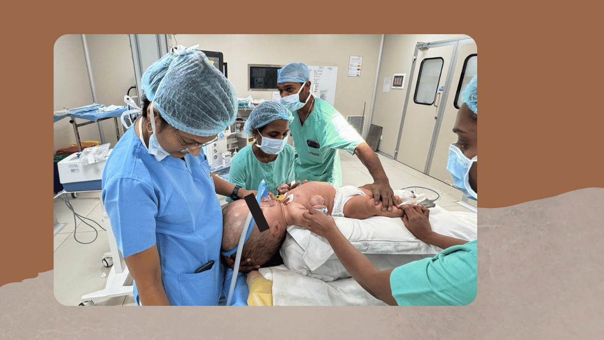
The level of consciousness, the presence of headache and vomiting, and the time course of symptom onset all inform how urgent the surgical intervention is and how carefully the anaesthetic induction must be managed to avoid acute neurological deterioration. In infants and young children, weight-based drug dosing, airway anatomy differences, temperature regulation, and smaller blood volumes all require specific attention. Having been mentored by internationally recognised paediatric anaesthesiologist Dr Rebecca Jacob and having participated in the Smile Train programme, paediatric neuroanaesthesia is an area of specific clinical experience in my practice.
Induction of anaesthesia: the most critical phase
The induction of anaesthesia in a patient with Raised intracranial pressure is the phase of highest risk. Laryngoscopy and intubation produce a transient but significant rise in blood pressure and heart rate that can translate into a dangerous spike in intracranial pressure. Managing this response requires precise timing and drug selection: intravenous agents that reduce intracranial pressure, opioids to blunt the haemodynamic response, and in some cases adjuncts such as lignocaine or antihypertensive agents.
Total intravenous anaesthesia using propofol-based techniques reduces cerebral metabolic rate and may be preferable to volatile inhalational agents in patients with significantly elevated intracranial pressure. The ventilation strategy during induction is equally critical: controlled ventilation targets a carbon dioxide level that maintains cerebral perfusion without causing cerebral vasoconstriction-related ischaemia.
Intraoperative management: maintaining cerebral homeostasis
Once anaesthetised and positioned for surgery, the focus of Neuro anaesthesia shifts to maintaining the conditions under which the neurosurgeon can work safely. This involves continuous monitoring through an invasive arterial line, end-tidal carbon dioxide as a proxy for arterial carbon dioxide levels, oxygen saturation, and in appropriate cases cerebral near-infrared spectroscopy.
The patient's head elevation improves venous drainage and reduces intracranial pressure, but excessive elevation can reduce cerebral perfusion. The neck must be positioned to avoid venous compression that would impede drainage. The osmotic agent mannitol is frequently administered to reduce brain swelling and provide better surgical conditions, requiring careful fluid balance management and monitoring of electrolytes.
Specific considerations for infant and neonatal VP shunt surgery
VP shunt surgery in the neonatal and infant period presents particular challenges absent in adult practice. The head of an infant with hydrocephalus may represent a disproportionately large fraction of total body weight, requiring specific positioning support. Temperature regulation is critical: neonates and small infants lose heat rapidly through the scalp and body surface, and hypothermia during surgery impairs coagulation and prolongs recovery. Awareness of potential VP shunt complications - including malposition, infection, and over-drainage - begins in the operative period, where anaesthetic decisions influence post-operative risk.
Neonates who have received gentamicin for sepsis, a common precipitant of post-haemorrhagic hydrocephalus in preterm infants, may have residual neuromuscular effects that influence the choice of muscle relaxant. Each of these factors is considered as part of the individual anaesthetic plan, and the team at Sparsh Hospital, Hennur, is experienced in the full range of paediatric neurosurgical cases.
Emergence from anaesthesia and post-operative neurological assessment
The emergence from anaesthesia after VP shunt surgery requires the same careful management as induction. A smooth, controlled emergence without coughing, straining, or haemodynamic instability is the goal since each of these can produce transient rises in intracranial pressure at a time when the surgical site is fresh. In Paediatric hydrocephalus cases, emergence assessment is particularly important since young children may not be able to communicate symptoms, making clinical observation — tone, responsiveness, fontanelle tension in infants - the primary tools for early complication detection.
Neurological assessment immediately after surgery is critical. The patient needs to be alert enough for a basic post-operative neurological check as soon as safely possible, so that any acute deterioration indicating a surgical complication can be identified promptly. This is one of the reasons why anaesthetic technique selection - specifically, using agents with rapid and predictable offset of action - matters in the recovery phase as much as induction.
Post-operative care and the role of the critical care team
After VP shunt surgery, patients are monitored in a neurosurgical high-dependency or intensive care setting where neurological observations are performed at regular intervals. The neuroanaesthesia and critical care team plays an active role: managing pain, monitoring for complications, and assessing the response to surgery. A functioning CSF shunt should produce a gradual improvement in symptoms - reduction in headache, improvement in alertness, and in infants a reduction in fontanelle tension and head circumference growth rate over subsequent weeks.
Complications to watch for in the immediate post-operative period include shunt malposition, subdural haematoma from over-drainage if the valve settings are too low, wound site infection, and systemic complications of anaesthesia. A sudden deterioration in the level of consciousness, a new focal neurological deficit, or worsening of headache in a patient who was improving should prompt urgent re-assessment and imaging.
Understanding brain fluid build-up after shunt insertion
One of the questions I hear most often from families after shunt surgery is whether the Brain fluid build-up will return and what signs to watch for. The shunt diverts CSF continuously, but it depends on the valve function remaining intact and the distal catheter in the peritoneal cavity remaining patent. Over time, the peritoneal end of the shunt can become blocked by omental adhesions, the ventricular catheter can become obstructed by choroid plexus ingrowth, or the valve can malfunction.
Signs of shunt malfunction mirror the original presentation of hydrocephalus: recurrent headaches, nausea, vomiting, lethargy, and in infants a bulging fontanelle or rapid head circumference growth. Any parent or patient who notices a return of symptoms that were previously controlled by the shunt should seek neurosurgical evaluation promptly rather than waiting for the next scheduled appointment.
Programmable valves and anaesthesia for future procedures
An important consideration for patients who have undergone Hydrocephalus surgery with a programmable valve is that subsequent exposure to strong magnetic fields, including MRI scanning, can alter the valve's pressure setting. Patients with programmable shunt valves should inform every anaesthesiologist, radiologist, and clinician of the valve type and current setting before any investigation or procedure. Before and after any MRI, the valve setting should be verified by the neurosurgical team.
In the anaesthetic context, this is particularly relevant when a patient with a pre-existing shunt presents for unrelated surgery. The neuroanaesthesiologist's pre-operative assessment should specifically enquire about valve type, current settings, and any recent symptoms of shunt malfunction, and coordinate with the neurosurgical team where any concern exists.
Questions families ask me most often
What is a programmable VP shunt and does my child need one?
Programmable VP shunt valves allow the neurosurgeon to adjust the pressure setting non-invasively using a magnetic device after surgery, without any further procedure. This flexibility is particularly valuable in children, where the optimal drainage pressure may need to be adjusted as the child grows, and in patients where over-drainage or under-drainage becomes apparent after the initial setting. Not every patient requires a programmable valve, and the neurosurgical team will discuss the most appropriate valve type for each individual based on diagnosis, age, and expected clinical course.
Will the anaesthesia affect my child's brain development?
The evidence on anaesthesia and neurodevelopment suggests that a single brief anaesthetic exposure in an otherwise healthy child carries very low risk of meaningful developmental impact. The risk of leaving hydrocephalus untreated, with ongoing pressure on developing brain tissue, is substantially greater than the risk of the anaesthetic required to treat it. I discuss this directly and honestly with every family, and I ensure that the anaesthetic technique and drug selection for each child minimises exposure duration and agent burden wherever clinically possible.
How long does the anaesthetic for shunt surgery last?
VP shunt insertion is typically a surgical procedure of 45 minutes to 1.5 hours, depending on whether this is a first insertion or a revision and any intraoperative findings. The anaesthetic begins before the surgical incision and continues through the closure. Recovery begins in the operating theatre immediately after surgery and continues in the recovery room, where the anaesthesia team remains present until the patient meets criteria for safe transfer.
Who should I see for a pre-anaesthetic consultation before shunt surgery?
A pre-anaesthetic consultation with a Neuroanaesthesiologist in Bengaluru who has specific experience in intracranial pressure management and paediatric neurosurgical cases is the most appropriate first step for families preparing for VP shunt surgery. This consultation reviews the imaging, clinical history, and any complicating factors, and allows the family to ask questions about the anaesthetic process before the day of surgery. At Sparsh Hospital, Hennur, I am available for pre-operative consultation for patients of all ages scheduled for VP shunt insertion or revision.
If your child or family member is scheduled for VP shunt surgery, or if you have questions about anaesthesia for a patient with existing hydrocephalus requiring another procedure, I am available for pre-operative consultation at Sparsh Hospital, HBR Layout, Hennur Road, Bengaluru 560043. To book a consultation with Dr Deepa Chandran, call 9686911560.
Written by Dr Deepa Chandran, MBBS (M S Ramaiah Medical College, Bengaluru), DA (Bangalore Medical College), DNB Anaesthesia (Bangalore Baptist Hospital), Fellowship in Ultrasound Guided Regional Anaesthesia (Jubilee Mission Hospital), Fellowship in Pain and Palliative Care, Consultant Neuroanaesthesiologist and Critical Care Specialist, Sparsh Hospital, HBR Layout, Hennur Road, Bengaluru 560043.
Related reading
Why Neuro Anaesthesia Is Different from General Anaesthesia
How Anesthesiologists Improve Outcomes in Complex Neurosurgeries
Inside the Neuro ICU: What Happens After Brain or Spine Surgery


Add a Comment