Skull base surgery is one of the most intricate and challenging fields within neurosurgery. It involves operating in the complex area where the brain, nerves, and blood vessels converge at the base of the skull. Over the years, I’ve dedicated myself to mastering these advanced techniques, as I believe that precision, detailed planning, and multidisciplinary teamwork are key to achieving successful outcomes for my patients.
Understanding the Significance of Skull Base Surgery
The skull base serves as the foundation for the brain and provides conduit pathways for nerves and blood vessels that control vital functions like vision, hearing, and facial sensation. Diseases such as tumors—meningiomas, chordomas, and schwannomas—or vascular abnormalities like aneurysms can develop in this delicate region. Proper surgical management not only eradicates these lesions but also preserves neurological function, balances risks, and improves quality of life.
Anatomical Complexity of the Skull Base
The anatomy of the skull base is incredibly intricate—with numerous tiny nerves and blood vessels running in close proximity. Structures like the optic nerves, carotid arteries, cranial nerves, and brainstem are situated just millimeters apart, making any surgical intervention extremely delicate. Miscalculations can lead to devastating consequences, including vision loss, paralysis, or life-threatening bleeding. This is why understanding the detailed anatomy and planning meticulously are critical steps in every case.
Preoperative Planning
Before I step into the operating theatre, I rely heavily on advanced imaging techniques such as MRI, CT scans, and angiography. These images help generate a detailed map of the tumor or lesion and its relationship to surrounding structures. For complex cases, I also utilize 3D navigation systems, which act as a GPS during surgery. This technology gives me real-time guidance, ensuring that every cut and maneuver is as precise as possible, minimizing risks to the patient.
Surgical Techniques
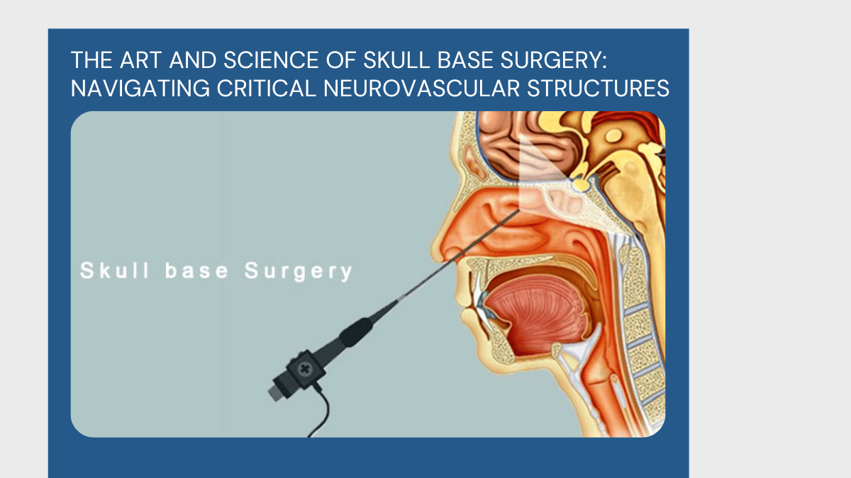
Skull base surgery offers various approaches depending on the lesion’s location, size, and accessibility. Endoscopic endonasal surgery—where we approach through the nose—is a revolutionary minimally invasive technique I frequently employ. It allows access to midline lesions with less tissue disruption. For lateral or more complex lesions, traditional transcranial approaches or combined methods are used. Each approach is tailored to the patient, ensuring safe removal while sparing vital structures.
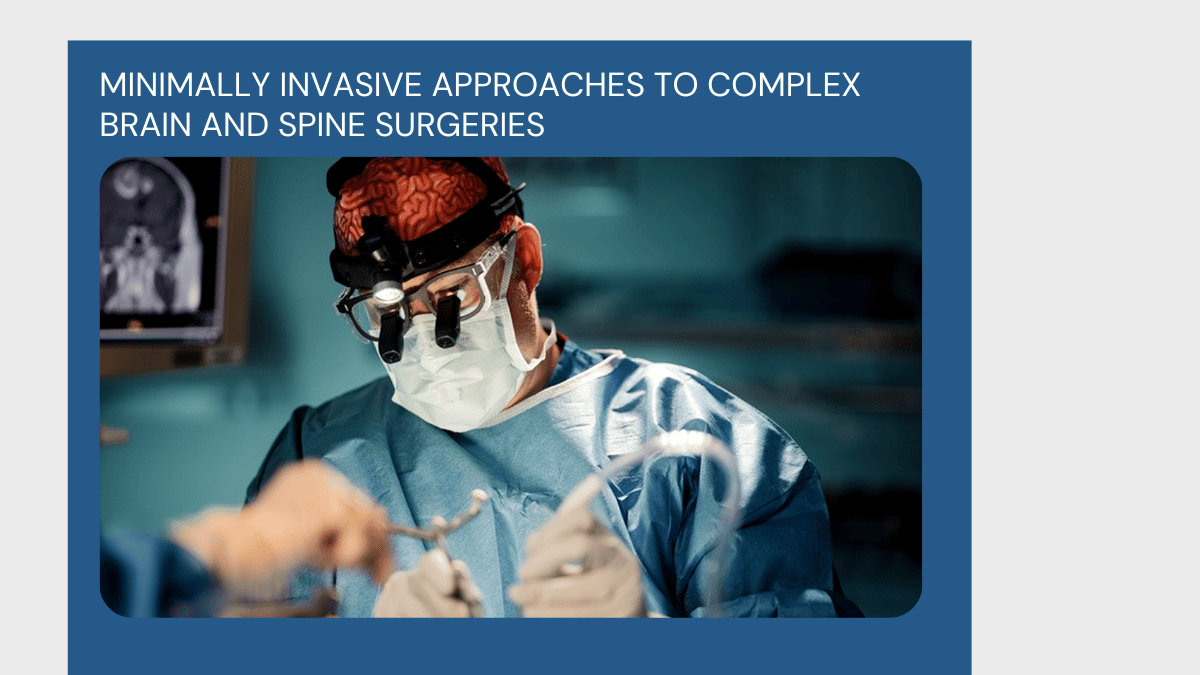
Intraoperative Precision: Tools and Techniques
During surgery, I harness advanced intraoperative tools such as neuronavigation, neuro-monitoring, and high-powered microscopes. These tools enhance visibility, provide real-time feedback, and help protect nerves and blood vessels. Neuro-monitoring alerts us immediately if a nerve is at risk, allowing us to adapt our approach instantly, greatly reducing postoperative complications.
Conditions Treated
My team and I have successfully treated numerous conditions, including meningiomas near the optic nerves, chordomas at the skull base, acoustic schwannomas affecting hearing and facial nerves, as well as vascular lesions like PICA aneurysms. Each case requires a customized strategy, combining meticulous planning and surgical expertise.
Postoperative Care and Rehabilitation
Postoperative care is crucial for optimal recovery. We adopt a multidisciplinary approach, involving neurologists, ophthalmologists, and physiotherapists, to ensure early rehabilitation and monitor for any complications. Close follow-up and imaging are performed to detect any residual or recurrent disease early.
Success Stories
One memorable case involved a patient with a large meningioma compressing the optic nerve. Using a minimally invasive endoscopic approach, we safely removed the tumor while preserving her vision. Today, she enjoys a normal life with no deficits—success that reflects the art and science of skull base surgery.
Moving Forward
With ongoing innovations like augmented reality, robotic assistance, and enhanced imaging, skull base surgery is becoming safer and more effective. As surgeons, our goal remains to combine the latest technology with our expertise—navigating this complex region with precision, compassion, and an unwavering commitment to our patients’ well-being.
Add a Comment