
Brain tumours are abnormal growths that develop in or around the brain. They may be benign or malignant, slow-growing or aggressive, primary or metastatic. Not all brain tumours are cancers. Some benign brain tumours, when safely and completely removed, may be cured with surgery and followed with periodic scans. The right plan depends on tumour type, location, symptoms, age, health, and molecular features.
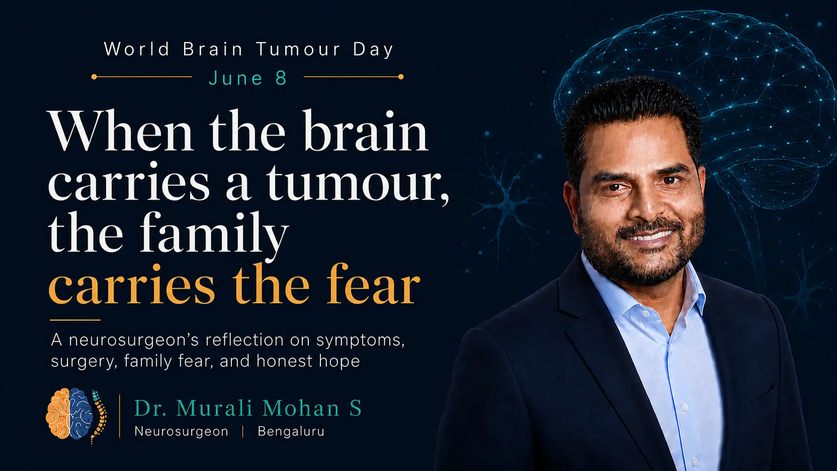
A brain tumour is one of the most frightening diagnoses a patient or family can hear. The fear is understandable. The brain is not just another organ. It carries movement, speech, vision, memory, behaviour, personality, emotion, and consciousness. Any disease that affects the brain immediately feels personal because it touches the very functions that make a person who they are.
But the word “brain tumour” is not a complete diagnosis by itself.
It is the beginning of a diagnostic journey.
Some brain tumours are discovered after seizures. Some after persistent headache. Some after vision problems, weakness, imbalance, speech difficulty, memory change, or personality change. Some are found incidentally during a scan performed for another reason. Some need urgent treatment. Some can be observed. Some are treated with surgery. Some need radiation, chemotherapy, targeted therapy, or a combination of treatments.
This guide is meant to help patients and families understand the subject in a structured way: what brain tumours are, why they occur, whether they are genetic, how they present, how they are evaluated, how prognosis is understood, what treatment now offers, and how life can continue after treatment.
What is a brain tumour?
A brain tumour is an abnormal growth of cells in the brain, its coverings, nearby nerves, the pituitary region, the skull base, or related structures.
Some brain tumours begin in the brain or central nervous system itself. These are called primary brain tumours. Others spread to the brain from cancers elsewhere in the body, such as lung, breast, kidney, colon, or melanoma. These are called metastatic brain tumours.
Brain tumours may be benign or malignant. This distinction is important because not all brain tumours are cancers. A benign brain tumour is non-cancerous and often grows slowly. In many patients, if a benign tumour can be safely and completely removed, surgery may be curative, and the patient may only need periodic follow-up scans afterward.
But benign does not always mean harmless. The brain sits inside the skull, and space is limited. Even a non-cancerous tumour can cause seizures, weakness, vision problems, hormonal disturbance, hydrocephalus, raised pressure, or compression of important nerves and brain areas depending on where it grows. So the question is not only whether the tumour is benign or malignant. The location, size, symptoms, growth pattern, and safety of removal matter just as much.
A malignant brain tumour grows more aggressively and may invade surrounding brain tissue. Some malignant tumours arise in the brain itself, while others are metastatic deposits from cancer elsewhere.
The brain is enclosed inside the skull. This means there is limited room for swelling, bleeding, pressure, or mass effect. Because of this, the location of a tumour can be as important as its size. A small tumour near the brainstem, optic nerve, speech area, or motor pathway may be more clinically significant than a larger tumour in a less sensitive region.
This is why the first question is not simply, “Is it cancer?”
The better questions are: what type of tumour is it, where is it located, what is it affecting, how fast is it growing, and what is the safest treatment plan?
Why do brain tumours occur?
Brain tumours occur when certain cells begin to grow in an abnormal and uncontrolled way.
In most patients, there is no single clear reason why the tumour developed. Brain tumours are usually the result of changes inside cells that affect how they grow, divide, repair damage, and interact with surrounding tissue. These changes may happen over time, and in many cases they are not linked to anything the patient did or failed to do.
This is important for families to understand. Patients often ask whether stress, diet, mobile phone use, old injuries, emotional trauma, or a particular lifestyle mistake caused the tumour. For most brain tumours, the honest answer is that there is no proven single cause that explains why it happened in that individual.
Some risk factors are known for certain tumours. Prior exposure to ionising radiation, especially therapeutic radiation to the head, is a recognised risk factor for some brain tumours. Certain inherited syndromes can increase risk. Metastatic brain tumours arise because cancer from another organ has spread to the brain.
But for many primary brain tumours, the cause remains unclear.
Medicine is improving its understanding tumour biology, especially through molecular testing. Today, many tumours are not classified only by how they look under the microscope. They are also classified by molecular changes inside the tumour cells. This helps doctors understand behaviour, prognosis, and treatment options more accurately.
The cause may not always be known. But the tumour can still be studied, classified, and treated in a planned way.
Are brain tumours familial or genetic?
Most brain tumours are not directly inherited from parents.
This is one of the most common fears families have. When one person is diagnosed, relatives may worry that children, siblings, or future generations are automatically at high risk. In most cases, that is not true.
There is a difference between a tumour having genetic changes and a tumour being inherited. Many brain tumours contain genetic or molecular changes within the tumour cells. These changes help the tumour grow and are useful for diagnosis and treatment planning. But these are usually acquired changes in the tumour itself, not inherited changes present in every cell of the body.
Familial brain tumours are uncommon. However, certain inherited syndromes can increase the risk of tumours in the brain and nervous system. Examples include neurofibromatosis, tuberous sclerosis, Li-Fraumeni syndrome, von Hippel-Lindau disease, and some other rare genetic conditions.
A genetic or familial evaluation may be considered when tumours occur at a young age, when there are multiple tumours, when there is a strong family history of related tumours, when there are associated skin or systemic features, or when the tumour type suggests an inherited syndrome.
For most patients, routine screening of all family members is not required. But if there is concern, the family should discuss it with the treating doctor, who may advise genetic counselling or further evaluation when appropriate.
Can environmental factors influence brain tumour risk?
The environmental influence on brain tumours is an area of active study, but for most patients there is no clear environmental explanation.
Ionising radiation is the most established environmental risk factor for certain brain tumours. This is usually relevant in people who previously received radiation therapy to the head, especially during childhood. Occupational, chemical, dietary, and lifestyle associations have been studied, but for most common brain tumours, the evidence is either limited, inconsistent, or not strong enough to blame a specific exposure in an individual patient.
Mobile phones are a common concern. Large studies and reviews have not established a clear causal link between regular mobile phone use and brain tumours, though research continues. Patients should avoid building guilt around everyday exposures unless there is strong medical evidence.
It is reasonable to live in a healthy way, avoid unnecessary radiation exposure, follow safety precautions in workplaces, and maintain general health. But patients should not carry the burden of thinking that a brain tumour happened because of one routine habit or one mistake.
In brain tumour care, guilt does not help. Understanding does.
What age do brain tumours occur?
Brain tumours can occur at any age.
They are seen in children, young adults, middle-aged people, and older adults. The type of tumour often varies with age. Some tumours are more common in children, while others are more common in adults or older patients.
In children, brain tumours may present with headache, vomiting, imbalance, vision problems, seizures, school decline, developmental concerns, or changes in behaviour. In very young children, symptoms can be difficult to recognise because they may not be able to describe what they feel.
In adults, brain tumours may present with seizures, headaches, weakness, speech changes, memory problems, personality change, vision symptoms, or symptoms related to cancer elsewhere in the body.
In older adults, symptoms may sometimes be mistaken for stroke, dementia, age-related weakness, depression, or general decline. A new seizure in an older adult, progressive neurological deficit, or unexplained cognitive change should be evaluated carefully.
Age matters because it influences the likely tumour type, treatment tolerance, surgical planning, rehabilitation needs, and goals of care. The same scan finding may be approached differently in a child, a young adult, and an elderly patient.
How do brain tumours present?
Brain tumours present according to their location, size, growth rate, swelling, pressure effect, and involvement of brain pathways.
A tumour in the motor area may cause weakness. A tumour near the speech area may cause difficulty speaking or understanding language. A tumour near the visual pathways may affect vision. A tumour in the cerebellum may cause imbalance or coordination problems. A tumour in the frontal lobe may cause personality, behaviour, judgement, or memory changes.
Some tumours cause seizures because they irritate the brain. In many adults, a first seizure may be the first sign of a brain tumour. Some tumours cause headache because of pressure, swelling, or blockage of cerebrospinal fluid pathways. Pituitary region tumours may cause hormonal symptoms, vision loss, menstrual changes, infertility, growth changes, weight changes, or sexual dysfunction.
Common presentations include:
- Persistent or worsening headache.
- Seizures, especially a first seizure in adulthood.
- Weakness or numbness of one side of the body.
- Difficulty speaking, understanding, reading, or writing.
- Vision problems, double vision, or loss of part of the visual field.
- Imbalance, dizziness, clumsiness, or walking difficulty.
- Memory change, personality change, confusion, or behavioural change.
- Vomiting, especially with headache or drowsiness.
- Hearing loss, ringing in one ear, or balance symptoms.
- Hormonal symptoms in tumours near the pituitary gland.
Many of these symptoms can occur due to conditions other than brain tumours. But persistent, progressive, new, or unusual neurological symptoms deserve medical evaluation.
How can brain tumours be recognised early?
Brain tumours are not always easy to recognise early because symptoms can be subtle, slow, or similar to common conditions.
A headache alone does not mean brain tumour. Most headaches are not due to tumours. But a headache that is new, progressively worsening, associated with vomiting, worse in the morning, accompanied by seizures, weakness, drowsiness, vision problems, or personality change should be evaluated.
A first seizure in an adult should always be investigated. Sudden weakness, speech difficulty, imbalance, or visual change may look like stroke and should be treated as urgent until evaluated. Gradual changes in personality, memory, work performance, or behaviour should not always be dismissed as stress, ageing, or emotional problems.
Early recognition is not about making people afraid of every symptom. It is about respecting patterns.
The warning pattern is usually one of change: a new symptom, a worsening symptom, a symptom that does not fit the person’s usual health, or a symptom associated with neurological signs.
When in doubt, the right step is not to compare on the internet. The right step is medical evaluation.
What are the types of brain tumours?
There are many types of brain and central nervous system tumours.
Gliomas arise from supporting cells of the brain. They include astrocytomas, oligodendrogliomas, and glioblastomas. Some gliomas grow slowly, while others behave aggressively. Molecular markers now play a major role in classifying gliomas and planning treatment.
Meningiomas arise from the coverings of the brain. Many are benign and slow-growing, but their location can make them important. A meningioma near the optic nerve, brainstem, major veins, or spinal cord may need a different approach from a small incidental meningioma.
Pituitary tumours arise near the base of the brain. They may affect hormones and vision. Some are treated with medicines, some with surgery, and some with observation or radiation.
Vestibular schwannomas, also called acoustic neuromas, arise from the balance and hearing nerve. They may cause hearing loss, tinnitus, imbalance, or pressure symptoms.
Medulloblastomas and other embryonal tumours are more often discussed in children, though some may occur in adults. Paediatric brain tumours have their own patterns, biology, and treatment pathways.
Metastatic brain tumours are tumours that have spread to the brain from cancer elsewhere in the body. Their treatment depends on the primary cancer, number of lesions, symptoms, systemic disease status, and available cancer therapies.
The American Brain Tumor Association notes that there are more than 120 types of primary brain and central nervous system tumours. This is why no two brain tumour stories should be compared too quickly. The word “tumour” is shared, but the biology may be completely different.
How do doctors prognosticate brain tumours?
Prognosis means estimating how a tumour may behave and what the expected course may be.
In brain tumour care, prognosis is not based on one factor alone. It depends on tumour type, grade, molecular markers, location, size, neurological condition, age, general health, extent of safe removal, response to radiation or chemotherapy, and whether the tumour is primary or metastatic.
A low-grade tumour may behave slowly but still need long-term follow-up. A high-grade tumour may need combined treatment and close surveillance. A benign tumour may be cured when it is completely and safely removed, but some benign tumours can recur, especially if a small part has to be left behind to protect important nerves or blood vessels.
A benign tumour may therefore have an excellent outlook in one patient and still require careful surveillance in another, depending on the tumour type and location. A malignant tumour may have more serious implications, but its treatment options and course depend on biology, molecular markers, response to treatment, and the patient’s overall condition.
Molecular diagnosis has changed prognostication. In gliomas, markers such as IDH mutation, 1p/19q codeletion, MGMT promoter methylation, and other molecular features can influence classification, treatment decisions, and expected behaviour. The 2021 WHO classification of central nervous system tumours gave molecular diagnostics a larger role in tumour classification.
Doctors should be honest about prognosis, but also careful. Prognosis is not a single sentence. It is a range of possibilities that becomes clearer as imaging, surgery, pathology, molecular reports, and treatment response are understood.
For families, the most useful question is often not “How long?” alone. It is also: what is the tumour type, what treatment is possible, what function can be preserved, what quality of life can be supported, and what should we prepare for?
What are the current promises in diagnosis?
Brain tumour diagnosis has improved greatly.
MRI remains the main imaging test for most brain tumours because it gives detailed information about the tumour’s location, size, relationship to important brain structures, swelling, enhancement pattern, and possible behaviour. Contrast MRI helps define the lesion better in many cases. CT scan remains useful in emergencies, especially when there is bleeding, hydrocephalus, raised pressure, trauma, or when MRI is not immediately available.
In selected patients, advanced MRI techniques add another layer of understanding. Diffusion imaging can help assess cellularity and tissue characteristics. Perfusion MRI can help estimate blood flow within and around the tumour. MR spectroscopy can provide metabolic information that may help distinguish tumour from infection, radiation change, or other mimics in selected situations.
When a tumour lies close to important functional areas of the brain, functional imaging becomes especially valuable. Functional MRI can help identify regions involved in movement, speech, or language before surgery. Tractography can help map important white matter pathways that carry motor, language, visual, and other signals. These tools help the surgical team understand not only where the tumour is, but also what important functions lie around it.
In selected cases, PET scan or SPECT imaging may also be used. These are functional imaging studies that look at metabolism or blood flow rather than anatomy alone. PET may help in selected brain tumours to assess tumour activity, distinguish recurrence from treatment-related change, or support planning when MRI findings are unclear. SPECT may be used in specific situations to understand blood flow or functional activity. These tests are not required for every patient, but they can be useful when the clinical question demands more than standard structural imaging.
Neuro-navigation helps surgeons use imaging as a guide during surgery. Awake brain mapping may be used in selected tumours near speech or language areas. Intraoperative ultrasound or intraoperative MRI may be used in some centres to assess tumour removal during surgery.
Pathology has also changed. Earlier, tumours were classified mainly by their microscope appearance. Today, molecular markers are increasingly important. The pathologist, molecular laboratory, and tumour board now play a central role in diagnosis.
This does not mean every patient needs every advanced test. It means the diagnosis is becoming more precise. Better diagnosis helps better planning.
The promise of modern diagnosis is not only seeing the tumour. It is understanding the tumour, its biology, its relationship to brain function, and the safest pathway for treatment.
What are the current promises in treatment?
Brain tumour treatment has moved from a single-modality approach to a more personalised, team-based model.
Surgery remains important for many tumours. It may remove the tumour, reduce pressure, improve symptoms, or provide tissue diagnosis. In selected benign tumours, complete safe removal may be the only active treatment needed apart from follow-up. In other tumours, surgery is one part of a larger pathway.
Radiation therapy has become more precise. Techniques such as stereotactic radiosurgery, fractionated radiotherapy, conformal planning, and proton therapy in selected settings allow radiation oncologists to target disease while limiting exposure to surrounding tissue as much as possible.
Chemotherapy and targeted therapy depend on tumour type. In some gliomas, molecular markers guide the use of chemotherapy. In metastatic brain tumours, systemic treatments, immunotherapy, and targeted medicines may influence brain disease management depending on the primary cancer.
Tumour boards bring multiple specialists together. A neurosurgeon, radiation oncologist, medical oncologist, radiologist, pathologist, neurologist, and rehabilitation team may all contribute to planning. This is especially important when decisions are complex.
The promise of modern treatment is not that every tumour can be cured. That would be false. The promise is that treatment can be more informed, more precise, more coordinated, and more tailored to the patient than before.
What is the role of surgery?
Brain tumour surgery has different goals in different patients.
Sometimes the goal is complete removal. In many benign brain tumours, if the tumour can be safely and completely removed and the patient recovers well, surgery may offer cure or long-term control. Examples may include selected meningiomas, schwannomas, and other benign tumours where complete excision is possible. These patients still need follow-up, because recurrence risk depends on tumour type, grade, location, and completeness of removal.
Sometimes the goal is safe maximum removal, where the surgeon removes as much tumour as possible while protecting speech, movement, vision, memory, and other critical functions. Sometimes the goal is biopsy, because diagnosis is needed before radiation, chemotherapy, or other treatment. Sometimes surgery is done urgently to reduce pressure, treat bleeding, or relieve hydrocephalus.
The central principle is safety.
This becomes especially important when a tumour is located in or near an eloquent area of the brain. Eloquent brain regions are areas that control important functions such as speech, movement, sensation, vision, memory, or understanding language. In such cases, the surgeon is not only thinking about tumour removal. The surgeon is also thinking about how to preserve the person’s ability to speak, move, see, understand, and live independently after surgery.
Modern neurosurgery has several tools that help in this planning. Functional MRI may help identify important areas related to movement or language before surgery. Tractography may help map important white matter pathways, such as motor, language, or visual tracts, so that the surgical route can be planned more carefully. Neuro-navigation allows the surgeon to use imaging guidance during the operation.
In selected patients, awake craniotomy may be used when a tumour is close to speech or language areas. During awake surgery, parts of the operation are performed while the patient is awake and responding, allowing the team to monitor speech, movement, or other functions in real time.
These tools do not remove all risk. They also do not apply to every patient or every tumour. But in carefully selected cases, functional MRI, tractography, awake craniotomy, intraoperative mapping, neuromonitoring, and neuro-navigation can improve surgical planning, help protect important brain functions, and support safer decision-making during surgery.
The surgeon must constantly balance tumour control with function preservation. A tumour near the speech area is different from one in the frontal pole. A tumour near the brainstem is different from one on the surface. A tumour in an elderly patient with other medical problems may need a different plan from a similar tumour in a young patient.
Modern neurosurgery uses microscopes, endoscopes, navigation, mapping, monitoring, ultrasonic aspirators, advanced anaesthesia, and neuro ICU care. These tools help, but they do not replace judgement.
A good surgical discussion should answer five questions clearly: why surgery is being advised, what it aims to achieve, what risks exist, what alternatives are available, and what recovery may look like.
What is life like after treatment?
Life after brain tumour treatment varies widely.
Some patients return to work, study, parenting, travel, and normal routines. Some need rehabilitation for weakness, balance, speech, swallowing, cognition, or daily activities. Some need anti-seizure medicines. Some need hormone replacement. Some need radiation or chemotherapy after surgery. Some need long-term surveillance scans. Some live with uncertainty around recurrence.
Recovery is physical, emotional, and social.
A patient may ask when they can drive, return to work, exercise, travel, or resume family responsibilities. A student may ask when they can return to school or college. A parent may worry about caregiving. A working adult may worry about income. An elderly patient may worry about independence.
These questions are not secondary. They are part of treatment.
Brain tumour follow-up should include wound care, medicines, seizure control, rehabilitation, pathology discussion, scan schedule, warning symptoms, and emotional support. In some patients, palliative care may also be important, not only at the end of life but earlier, to improve comfort, decision-making, and support.
The goal is not only to treat the tumour. The goal is to help the person live as fully and safely as possible.
Brain tumour survivors: where hope lives
Brain tumour survivorship is a growing and important part of care.
A survivor may be someone who has completed treatment and is on follow-up. It may be someone living with a stable tumour. It may be someone undergoing long-term therapy. It may be someone adapting to new limitations after surgery, radiation, or chemotherapy.
Survivorship is not always simple. Patients may fear every follow-up scan. They may experience fatigue, seizures, memory difficulty, weakness, mood changes, sleep problems, or anxiety. Families may continue to live with uncertainty even after treatment appears successful.
For patients with benign tumours that have been completely removed, survivorship may involve returning to normal life while continuing periodic surveillance. For patients with malignant or recurrent tumours, survivorship may involve ongoing treatment, rehabilitation, symptom control, and emotional support.
Hope in survivorship is not denial. It is structure.
Hope is follow-up. Hope is rehabilitation. Hope is seizure control. Hope is returning to school, work, family, and social life where possible. Hope is knowing what symptoms to watch for. Hope is having a team that continues to guide the patient after the first treatment is over.
Many brain tumour survivors live meaningful lives after diagnosis and treatment. Their journeys may be changed, but not erased. This is an important message for newly diagnosed patients: the diagnosis is serious, but it is not the whole of a person's identity.
The scan shows disease. Life still contains the person.
Why teamwork matters
Brain tumour care is strongest when it is team-based.
The team may include a neurosurgeon, neurologist, radiologist, pathologist, radiation oncologist, medical oncologist, endocrinologist, neuro-anaesthetist, intensivist, physiotherapist, speech therapist, occupational therapist, psychologist, nurse, social worker, and palliative care specialist.
Not every patient needs every specialist. But complex brain tumour care often needs multiple perspectives.
The neurosurgeon may remove tumour or obtain tissue. The radiologist helps interpret imaging. The pathologist defines the tumour. The radiation oncologist plans radiation. The medical oncologist guides chemotherapy or systemic treatment. The neurologist helps manage seizures and neurological symptoms. Rehabilitation specialists help the patient regain function. Nurses and caregivers support daily recovery.
The patient is at the centre. Around the patient is the team.
This is how brain tumour care becomes more than an operation, a scan, or a report. It becomes a pathway.
What should patients and families remember?
Patients and families should remember that a brain tumour is not one disease and should not be understood through fear alone.
The important questions are: what type of tumour is it, where is it, what is it affecting, what does the pathology show, what do the molecular markers suggest, what treatment is possible, what function must be protected, and what follow-up is needed?
Not every brain tumour is cancer. Not every tumour needs immediate surgery. Not every tumour can or should be completely removed. Some benign tumours, when safely and completely removed, may be cured with surgery and followed with scans. Other tumours need longer treatment and closer monitoring. Not every recurrence means nothing can be done. Not every online story applies to every patient.
The first step is careful evaluation.
The next step is a clear plan.
A brain tumour diagnosis changes life, but it does not remove the need for clarity, dignity, and hope. The best care is scientific enough to understand the tumour and humane enough to see the person living with it.
This article is for awareness and education. It is not a substitute for medical advice. Anyone with new seizures, persistent or worsening headaches, weakness, speech difficulty, vision change, confusion, drowsiness, imbalance, or other concerning neurological symptoms should seek in-person medical evaluation.


Add a Comment