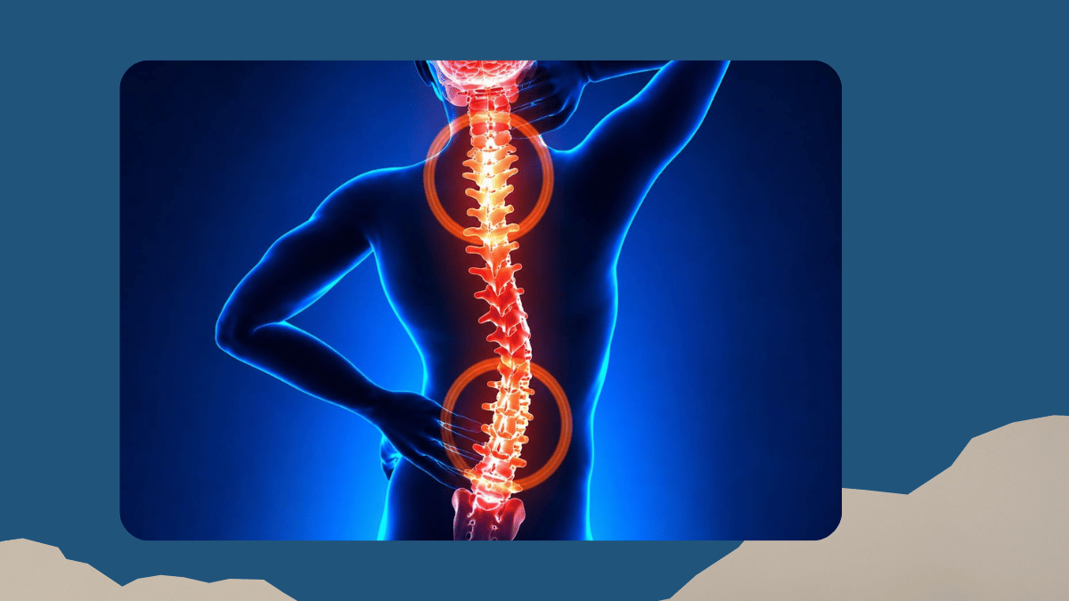
When someone is told they need to see a neurosurgeon, the first response is almost always a mixture of anxiety and a flood of questions: what does a neurosurgeon actually do, what conditions require surgery, how do you know if surgery is the right option, and what does recovery look like? As a neurosurgeon in Chennai with more than 25 years of experience and over 8,000 neurosurgical procedures performed at Gleneagles Fortis Hospital, Perumbakkam, I want to answer all of those questions clearly, in one place, for every patient and family who needs them.
What does a neurosurgeon treat?
Neurosurgery is the speciality concerned with the surgical treatment of conditions affecting the brain, spinal cord, spine, peripheral nerves, and related vascular structures in the brain. Most people associate neurosurgery with brain tumour operations, and that is certainly a significant part of the speciality. But the scope of neurosurgery is considerably broader, encompassing conditions of the spine, the cerebrospinal fluid system, movement disorders, epilepsy, cerebrovascular disease including aneurysms and arteriovenous malformations, skull base pathology, and neurological conditions in children. A neurosurgeon is not simply someone who operates on the brain: they are a specialist in the full spectrum of nervous system surgery from the top of the skull to the lower end of the spine.
Brain disorders that require neurosurgical treatment
Brain tumours
Brain tumours are among the conditions I treat most frequently in my practice. They range from benign, slow-growing lesions that cause symptoms through pressure on surrounding structures, to malignant tumours that require prompt and aggressive management. The distinction between benign and malignant tumours matters, also location matters as much as pathology: a small benign tumour in a critical area of the brain can be more challenging to manage than a larger tumour in a more accessible location. Modern brain tumour surgery uses neuronavigation, intraoperative monitoring, and when appropriate, awake craniotomy techniques to maximise safe resection while preserving neurological function. I have published extensively on colloid cysts, meningiomas, and paediatric brain tumours, and my practice includes the full range of cranial tumour pathology.
Skull base surgery
The skull base is the most anatomically complex region of the head, where the brain sits on a shelf of bone perforated by critical nerves and blood vessels. Tumours and lesions in this region require specialised approaches that access the pathology while respecting these critical structures. Skull base surgery includes approaches to pituitary tumours, acoustic neuromas, meningiomas of the skull base, chordomas, and other lesions in this demanding anatomical region. My fellowship experience in complex skull base surgery in Australia, combined with 25 years of operative experience, informs how I approach these cases at Gleneagles Fortis Hospital, Chennai.
Hydrocephalus and CSF disorders
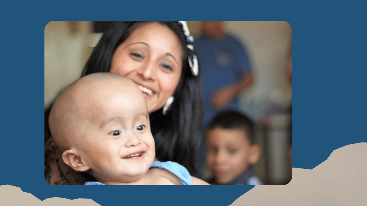
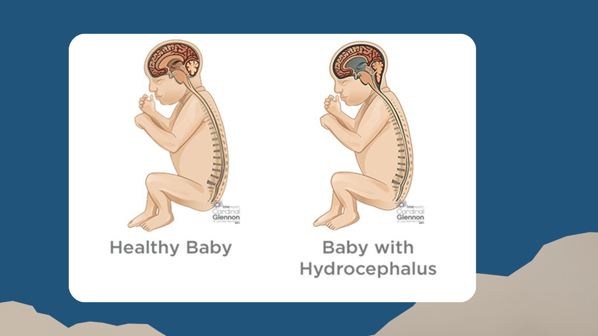
Hydrocephalus is a condition in which cerebrospinal fluid accumulates in the brain's ventricular system, causing progressive pressure on brain tissue. It can affect newborns, children, and adults, and its causes and optimal treatment vary significantly by age and aetiology. I have been a member of the Hydrocephalus Research World Record Ranking Committee and have published multiple papers in international journals on hydrocephalus management and shunt systems. Hydrocephalus treatment in appropriate cases uses endoscopic third ventriculostomy (ETV), which avoids the need for a permanent shunt by creating a natural drainage pathway within the brain itself, and is one of my areas of particular expertise.
Cerebrovascular conditions
Cerebrovascular surgery addresses conditions of the blood vessels supplying the brain. Intracranial aneurysms, which are balloon-like weaknesses in arterial walls that can rupture and cause life-threatening subarachnoid haemorrhage (SAH), require either microsurgical clipping or endovascular coiling depending on their size, shape, and location. Arteriovenous malformations (AVM) are tangles of abnormal blood vessels that can cause haemorrhage, seizures, or progressive neurological deficit. Cerebrovascular surgery in Chennai for these conditions requires a combination of microsurgical skill and precise pre-operative planning using modern cerebrovascular imaging, and forms an important part of my neurosurgical practice.
Traumatic brain injury
Traumatic brain injury encompasses the full spectrum of brain damage caused by external force, from mild concussion to severe diffuse axonal injury requiring intensive neurosurgical management. The neurosurgical role in traumatic brain injury includes evacuation of haematomas that are causing dangerous pressure on the brain, decompressive craniectomy to relieve refractory intracranial hypertension, and the ongoing management of intracranial pressure in the neurosurgical intensive care environment. Timing is critical in traumatic neurosurgery: the outcome of an expanding haematoma is dramatically better when surgical decompression is achieved within the appropriate window.
Spine disorders requiring neurosurgical treatment
Degenerative spine disease
Degenerative spine disease is one of the most common sources of neurological disability in adults, and encompasses conditions including cervical spondylosis, lumbar disc prolapse, spinal stenosis, and degenerative instability requiring fusion. The fundamental question in spine surgery is whether the patient's symptoms, the imaging findings, and the failure of appropriate non-surgical management together justify an operative intervention. Not every disc prolapse needs surgery; not every patient with spondylosis is a surgical candidate. When surgery is indicated, minimally invasive spine surgery using tubular retractors, endoscopic access, and microincisions reduces tissue disruption, shortens recovery, and achieves comparable or superior outcomes to traditional open surgery in appropriate cases. My spinal fellowship at Flinders Medical Centre, Adelaide, specifically trained me in these techniques.
Spinal tumours
Spinal tumours may arise within the spinal cord itself, from the covering membranes, or from the bony vertebral column. Intradural tumours, those inside the dural covering, include meningiomas, schwannomas, and ependymomas, and are generally amenable to surgical resection with good outcomes when approached carefully. Extradural tumours include metastatic deposits from primary cancers elsewhere in the body and primary bone tumours of the spine. The management of each type requires individual evaluation of the tumour's pathology, location, and the patient's overall health and prognosis.
Spinal trauma
Spinal injuries from road accidents, falls, and other high-energy mechanisms represent a neurosurgical emergency when they cause instability or neurological compromise. The decision to operate involves assessing the stability of the injury, the degree of neurological deficit, and the likelihood of recovery with or without surgical decompression and stabilisation. Spinal reconstruction and fusion in trauma uses modern implant systems to restore spinal column stability, protect the spinal cord from further injury, and allow early mobilisation, which significantly improves rehabilitation outcomes compared to prolonged immobilisation.
Functional neurosurgery: treating the brain's circuits
Functional neurosurgery is the subspeciality focused not on removing pathological tissue but on modifying the function of specific brain circuits to treat neurological and psychiatric conditions. The most established functional procedure is deep brain stimulation (DBS), in which electrodes are precisely implanted into target structures deep in the brain and connected to a pulse generator (battery) beneath the skin. Electrical stimulation through these electrodes modulates abnormal neural activity, providing sustained symptom relief for Parkinson's disease, essential tremor, dystonia, and in selected cases of obsessive-compulsive disorder.
DBS surgery requires millimetre-level precision in electrode placement, achieved through stereotactic targeting using preoperative MRI and CT, planning station, intraoperative physiological mapping, and in appropriate cases, awake surgery so that the patient's response to stimulation can be assessed in real time during the procedure. My functional neurosurgery fellowship at Flinders Medical Centre gave me specific training in these techniques, and functional neurosurgery is one of the central pillars of my practice at Gleneagles Fortis Hospital, Chennai.
Paediatric neurosurgery: treating neurological conditions in children
Neurological conditions in children present with different patterns, different causes, and different technical requirements compared to the same conditions in adults. Paediatric neurosurgery requires adaptations of technique, instruments, and anaesthetic approach to the smaller anatomy and different physiology of children, and an understanding of the developmental implications of both the condition and the treatment. My fellowship in paediatric neurosurgery at Jikei University School of Medicine, Tokyo, under Oi-Sami Fellowship, with Prof Oi being one of the world leaders in paediatric hydrocephalus and neuroendoscopy, provided specific training in this area.
Paediatric neurosurgical conditions I treat include hydrocephalus from multiple causes including post-haemorrhagic, congenital, and tumour-related; Moyamoya disease, a rare cerebrovascular condition causing progressive stroke in children that can be treated with indirect bypass surgery; paediatric brain tumours including medulloblastoma, ependymoma, astrocytoma, and choroid plexus tumours; congenital malformations of the brain and spine; and drug-resistant epilepsy amenable to surgical resection or disconnection.
Neuroendoscopy: minimally invasive neurosurgery from within
Neuroendoscopy uses a thin telescope introduced through a small burr hole in the skull to visualise and treat conditions within the brain's ventricular system. Neuroendoscopy avoids the need for a large craniotomy in appropriate cases and is particularly valuable for the treatment of intraventricular tumours, colloid cysts of the third ventricle, and hydrocephalus. I have published extensively on the transcallosal transforaminal approach to colloid cysts, reporting a 20-year experience in the Neurology India journal, and my training in neuroendoscopy in Japan gives me specific expertise in this area. Endoscopic third ventriculostomy, the endoscopic treatment for certain types of obstructive hydrocephalus, is one of the most effective procedures in my armamentarium for selected patients.
Epilepsy surgery: when medication is not enough
Drug-resistant epilepsy, defined as failure of adequate trials of two appropriately chosen antiepileptic drugs, affects approximately one third of people with epilepsy and represents a significant quality-of-life burden. Epilepsy surgery offers the possibility of meaningful seizure reduction or complete seizure freedom in carefully selected patients through resection of the seizure-generating zone, disconnection procedures, or neuromodulation approaches such as vagus nerve stimulation or responsive neurostimulation. The Advanced Epilepsy Centre at Gleneagles Fortis Hospital, Chennai is equipped with 3 Tesla MRI, PET scan, video EEG, and Stereo EEG for pre-surgical evaluation of complex cases, and in the last six months I have performed epilepsy surgery successfully in many patients with drug-resistant epilepsy using this comprehensive evaluation pathway.
Modern technologies in neurosurgery
The quality and safety of neurosurgical care is directly dependent on the technology available to the surgeon. Several technologies have transformed what is possible in modern neurosurgery. Neuronavigation uses preoperative MRI and CT data merged in real time with the patient's actual position on the operating table, allowing the surgeon to navigate to targets with sub-millimetre accuracy. Intraoperative neurophysiological monitoring tracks the function of critical neural pathways during surgery, providing real-time feedback that allows the surgeon to adjust technique and reduce the risk of neurological deficit. High-powered operating microscopes provide magnification and illumination that allow surgical dissection at a cellular level. Intraoperative imaging, including intraoperative MRI and ultrasound, allows the surgeon to assess the extent of tumour resection while the patient is still on the table and address residual tumour before wound closure.
Recovery after neurosurgery: what patients should expect
Recovery from neurosurgical procedures varies enormously depending on the procedure, the underlying condition, and the patient's pre-operative neurological status. Minimally invasive spine surgery patients often go home within one to two days and return to light activity within two to four weeks. Cranial surgery for tumours or vascular conditions typically involves a hospital stay of five to eight days, with recovery continuing at home over weeks to months. The most important determinant of long-term outcome is the underlying condition and the completeness of surgical treatment, not the surgery itself. Neurosurgery recovery in my practice is supported by close postoperative follow-up, physiotherapy where indicated, and coordination with neurology, neuro-oncology, and rehabilitation medicine as the clinical picture requires.
Questions patients ask me most often
How do I know if my symptoms need a neurosurgical evaluation?
The decision to see a neurosurgeon does not mean that surgery is inevitable or even likely. It means that your symptoms have a neurological basis that warrants specialist evaluation. Persistent headaches that are new in pattern or progressive, limb weakness or numbness, back pain radiating into the legs with neurological deficit, a first seizure in adult life, balance problems of progressive onset, or an abnormal finding on brain or spine imaging are all reasons to see a neurosurgeon. The neurosurgical evaluation determines whether the condition is surgical, and if so, what surgical approach is optimal.
Is neurosurgery always high-risk?
All surgery carries risk, and neurosurgery on the brain and spinal cord carries specific risks related to the structures involved. However, the risk profile of modern neurosurgery is significantly lower than it was two decades ago because of improvements in imaging, navigation, intraoperative monitoring, anaesthesia, and minimally invasive techniques. The question for any neurosurgical procedure is not whether there is risk, but whether the risk of surgery is lower than the risk of the natural history of the untreated condition. For many conditions, including certain brain tumours, hydrocephalus, and ruptured aneurysms, neurosurgical risk without surgery is substantially higher than the risk of the operation itself. This is the context within which every surgical decision is made.
What should I bring to my first neurosurgical consultation?
Bring all existing imaging: MRI and CT discs and reports, not just printed images. Bring a written summary of your symptoms including when they started, how they have changed, and what makes them better or worse. Bring a list of all current medications. If you have had previous neurosurgical procedures or neurological investigations, bring the operative notes and reports where possible. The more complete the clinical picture at the first consultation, the more productive the appointment will be for both of us.
Can I get a second opinion before deciding on surgery?
Absolutely, and I actively encourage it. A significant neurosurgical decision deserves thorough consideration, and any surgeon who discourages a second opinion is not serving the patient's best interest. My role is to give you the best clinical assessment I can, explain what I recommend and why, and support whatever decision you make. Second opinions for complex neurosurgical conditions, particularly brain tumours, cerebrovascular conditions, or spine surgery, are entirely appropriate and often valuable.
If you have been referred to a neurosurgeon, are considering seeking an evaluation for neurological symptoms, or would like a second opinion on a neurosurgical recommendation you have received, I am available at Gleneagles Fortis Hospital, Perumbakkam, Chennai.
Written by Dr Nigel Peter Symss, MBBS, DNB (Neurosurgery), Fellowship Paediatric Neurosurgery (Jikei University, Japan), Fellowship Spinal, Functional Neurosurgery and Microneurosurgery (Flinders Medical Centre, Australia), HOD and Senior Consultant Neurosurgeon and Functional Neurosurgeon, Gleneagles Fortis Hospital, Perumbakkam, Chennai.
Related reading
Brain Tumours: Symptoms, Diagnosis, and How Modern Surgery Has Changed Outcomes
Epilepsy and Epilepsy Surgery: What You Need to Know
Understanding Hydrocephalus in Children: Causes, Symptoms, and Treatment
Deep Brain Stimulation A Life Changing Option for Parkinson’s Patients

Comments
Dr Murali Mohan
Posted on: August 11,2026 Published
Wonderful Article Dr Nigel, Keep writing Keep enlightening 💐
Add a Comment