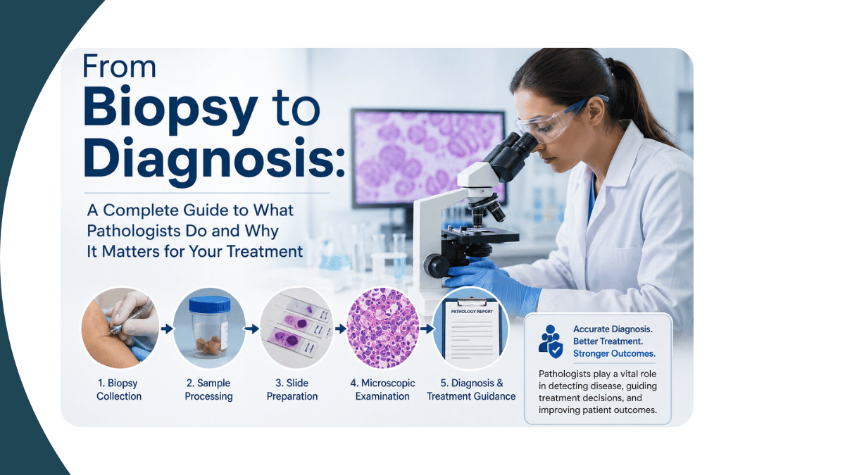
When a patient is told that they need a biopsy, or that their tissue has been sent to pathology, the natural question that follows is: what exactly is the pathologist doing with it, and why does the pathologist's report determine what treatment I receive? The pathology report is the document that defines your diagnosis. It is the basis on which your surgeon decides whether to operate, your oncologist decides which chemotherapy to prescribe, and your treating physician decides whether your condition is inflammatory, infectious, benign, or malignant. As a Professor and Head of the Department of Pathology at PES Institute of Medical Sciences and Research, Kuppam, with more than 20 years of diagnostic pathology experience and 75 peer-reviewed publications, I want to explain the full landscape of diagnostic pathology, what each investigation involves, what the report means, and how to use pathology to make the most informed decisions about your health. Pathology is not a supporting service in medicine; it is the diagnostic foundation on which every treatment decision in cancer, inflammation, and organ disease is built.
What Does a Pathologist Do?
The pathologist is the physician who examines tissues, cells, and body fluids under the microscope to produce a diagnosis. Unlike the clinician who assesses symptoms and examines the patient, or the radiologist who interprets imaging, the pathologist examines the actual cellular architecture of diseased tissue to determine what is happening at the microscopic level. This cellular analysis is the most definitive form of medical diagnosis available. A mass on a CT scan can be described in terms of its size, location, and imaging characteristics. A pathology report describes exactly what type of cells it is made of, whether they are malignant, how aggressive they appear, and which proteins they express on their surface, information that directly determines which targeted therapy will work against them. In cancer diagnosis, the pathology report is never just a confirmation of what imaging suspected; it is a precise molecular characterisation of the specific cancer type that guides individualised treatment.
The pathologist works across several disciplines: histopathology (tissue examination), cytopathology (cell examination from body fluids or aspirates), hematopathology (blood and bone marrow disorders), clinical pathology (laboratory investigation of body fluids), and immunohistochemistry (protein expression analysis on tissue). Each serves a different diagnostic function, and in complex cases, several of these are combined to reach the final diagnosis.
Fine Needle Aspiration Cytology (FNAC): The First Investigation for Lumps and Swellings
Fine needle aspiration cytology, universally known as FNAC, is one of the most valuable and most accessible diagnostic tools in pathology. It involves passing a fine needle into a palpable lump, whether in the thyroid, lymph node, breast, salivary gland, soft tissue, or any other accessible structure, and aspirating a small amount of material. This material contains cells from the lesion, which are spread on glass slides, stained, and examined under the microscope.
FNAC provides a rapid, minimally invasive preliminary assessment of whether a lump is benign, inflammatory, or malignant. For a thyroid nodule, FNAC can distinguish a benign colloid nodule from a follicular neoplasm from a papillary carcinoma within days. For a neck lymph node, FNAC can identify whether it is reactive (from infection), granulomatous (from tuberculosis), or metastatic cancer. For a breast lump, FNAC provides a same-session preliminary assessment that guides the management conversation before the patient leaves the clinic. Ultrasound-guided FNAC, which uses real-time imaging to direct the needle precisely into the target lesion, has significantly improved the accuracy of cytological sampling for deep or small lesions, and at PESIMSR, Kuppam, this technique is routinely applied for lesions that are not palpable at the surface. FNAC does not replace histopathology for definitive cancer diagnosis, but it is the most efficient and most patient-friendly first step.
Histopathology: The Gold Standard for Tissue Diagnosis
If FNAC is the first step, histopathology is the definitive step. Histopathology involves the examination of a tissue biopsy or a surgically removed specimen under the microscope. The tissue is fixed in formalin, embedded in paraffin wax, cut into extremely thin sections (typically four to five micrometres thick, thinner than a single cell diameter), mounted on glass slides, and stained with haematoxylin and eosin (H&E), the standard stain that makes cell nuclei appear blue and cytoplasm pink under the microscope.
The pathologist examines the tissue architecture, the relationship between cells, the type and character of the cells present, the presence of invasion, the state of the surgical margins (whether the tumour has been completely removed or whether cancer cells are present at the cut edges), and the extent of spread to lymph nodes or adjacent structures. This examination produces the information that defines the cancer diagnosis completely: its type, grade, stage, lymphovascular invasion status, perineural invasion, margin status, and lymph node involvement.
The histopathology report is the document that the multidisciplinary tumour board reviews when deciding whether a patient needs additional surgery, radiotherapy, chemotherapy, or targeted therapy. The pathologist who signs out this report is not performing a clerical function. Each histopathology report represents a diagnostic judgement that requires the integration of cellular morphology, clinical context, imaging findings, and subspecialty expertise to produce an accurate and clinically useful result, and the accuracy of that report is the most important single factor determining whether the right treatment is given.
Immunohistochemistry: Identifying What Cells Express
Immunohistochemistry (IHC) is a technique performed on tissue sections that uses specific antibodies to detect the presence or absence of particular proteins within cells and tissues. In cancer pathology, IHC has transformed diagnosis and treatment selection. It allows the pathologist to answer questions that H&E staining alone cannot: Is this breast cancer hormone receptor positive? Does this lung cancer overexpress HER2? Is this lymphoma a B-cell or T-cell type? Are these cells expressing a specific protein that a targeted therapy can attack?
In breast cancer, IHC for oestrogen receptor (ER), progesterone receptor (PR), and HER2 determines whether a patient is a candidate for hormone therapy, anti-HER2 therapy (trastuzumab), or chemotherapy only. In lymphomas, a panel of IHC markers defines the exact subtype, which determines the chemotherapy regimen and the prognosis. In soft tissue tumours, where many different tumour types can look similar on H&E, IHC is essential for distinguishing between entities that require completely different treatments. I have extensive experience in designing and interpreting IHC panels at PESIMSR, Kuppam, and my research includes publications on tumour budding, stromal tumour-infiltrating lymphocytes, and post-chemotherapy histopathological changes in breast carcinoma, all of which rely on precise IHC interpretation. IHC bridges the gap between morphological diagnosis and molecular treatment selection, making it the most important technical advance in surgical pathology of the last three decades.
Cytopathology, Hematopathology, and Specialist Pathology Areas
Beyond FNAC and tissue biopsy, the pathology laboratory provides diagnostic services across a wide clinical spectrum.
Cervical Cytology and Cancer Screening
The cervical smear (Pap smear) examination is one of the most important cancer screening tests available, capable of detecting pre-cancerous cervical changes years before invasive cancer develops. I hold a certification in Cervical Cancer Screening for Pathologists from the ICMR-National Institute of Cancer Prevention and Research (NICPR), and cervical cytology is a significant part of our diagnostic volume at PESIMSR, Kuppam, where access to this screening for women in Chittoor district and the surrounding Andhra Pradesh region is a clinical priority.
Hematopathology: Blood Disorders and Bone Marrow
Hematopathology covers the diagnosis of blood disorders including anaemia, clotting disorders, and blood cancers including leukaemia, lymphoma, and myeloma. Blood film examination, bone marrow aspirate, and trephine biopsy are the principal investigations. My published work on coagulation parameters in COVID-19, cytomorphological patterns in lymph node aspirates, and adolescent menorrhagia coagulation profiles reflects the breadth of hematopathological work at a teaching hospital like PESIMSR. Hematological malignancies in particular require a combination of morphology, IHC, flow cytometry, and cytogenetic testing to reach a definitive diagnosis and treatment plan , a complexity that makes experienced hematopathology essential for patients presenting with blood abnormalities or suspected haematological cancers.
Dermatopathology, Nephropathology, and Other Subspecialties
Subspecialty pathology areas require specific training beyond general histopathology. Dermatopathology involves the interpretation of skin biopsies for inflammatory skin conditions, autoimmune diseases, and skin cancers. My published work includes Tzanck smear cytology in dermatology, mast cell distribution in skin disorders, and psoriasis immunohistochemistry, reflecting active clinical and research involvement in dermatopathology. Nephropathology, the interpretation of kidney biopsies for glomerular and tubular diseases, requires a combination of light microscopy, immunofluorescence, and electron microscopy, and I hold an ISN-ANIO Basic Clinical Nephropathology Certificate Programme qualification. Placental and perinatal pathology, including examination of placentas in pre-eclampsia, eclampsia, and fetal growth restriction, is another area in which I have active publications, including a scoring system for placental pathology in pre-eclampsia.
Understanding Your Pathology Report: What the Key Terms Mean
Pathology reports are written for other clinicians and are not designed to be immediately understandable to patients, but several terms recur across reports and understanding them helps patients engage with their diagnosis and ask the right questions at the next clinical appointment. Grade refers to how abnormal the cancer cells look under the microscope compared to normal cells: low-grade cancers resemble normal cells more closely and usually grow more slowly, while high-grade cancers are more abnormal and typically more aggressive. Stage refers to the extent of spread of the cancer: stage I disease is localised, stage IV disease has spread to distant organs. Margins refer to the edges of the surgical specimen: clear margins (also called negative or free margins) mean that no cancer cells are at the cut edges, while involved margins (positive margins) mean that cancer extends to the resection edge and that further surgery or radiotherapy may be needed. Lymphovascular invasion means that cancer cells have been found inside small blood vessels or lymph channels within the specimen, which increases the risk of spread. Lymph node status, the number of lymph nodes examined and the number involved by cancer, directly determines staging and systemic treatment decisions. Reading a pathology report together with your treating doctor and asking specifically what each of these findings means for your treatment plan is the most productive conversation a patient can have after a cancer diagnosis.
When to Seek a Pathology Second Opinion
A pathology second opinion involves sending the original tissue slides or blocks to a second pathologist for independent review. It is not an expression of distrust in the original pathologist. It is a clinical safeguard that is standard practice in many countries and that is particularly important in specific situations. Second opinions are most valuable when the diagnosis is rare or unusual, when the tumour type falls outside the routine experience of the original laboratory, when the report is inconclusive or describes an atypical presentation, when the proposed treatment is aggressive and irreversible (such as major surgery or bone marrow transplantation), and when there is any discordance between the clinical impression and the pathology finding. At PESIMSR, Kuppam, I provide pathology consultations and second opinions for cases referred from across the Chittoor district and the surrounding region. My subspecialty experience in breast pathology, gynaecological pathology, dermatopathology, and nephropathology makes me particularly well placed to review cases in these areas. My 75 peer-reviewed publications, spanning histopathology, cytopathology, IHC, hematopathology, dermatopathology, and nephropathology, reflect the breadth of diagnostic pathology that I bring to complex case review. If you have received a pathology report and are uncertain about the diagnosis or its implications for your treatment, a formal second opinion consultation is the most appropriate next step.
To reach Dr. Anikode Subramanian Ramaswamy for a pathology consultation or second opinion at PES Institute of Medical Sciences and Research (PESIMSR), NH-219, Kuppam, Nalagampalle, Andhra Pradesh 517425, call +91 9493726494.
Written by Dr. Anikode Subramanian Ramaswamy, MBBS, MD (Pathology), Professor and Head, Department of Pathology, PES Institute of Medical Sciences and Research (PESIMSR), NH-219, Kuppam, Nalagampalle, Andhra Pradesh 517425. ISO 15189 Assessor, NABH Internal Counsellor. 75 peer-reviewed publications. Phone: +91 9493726494.
Related reading
Histopathology and Cancer Diagnosis: How Tissue Analysis Guides Treatment Decisions
Why Quality Management Is the Foundation of Safe and Effective Healthcare


Add a Comment