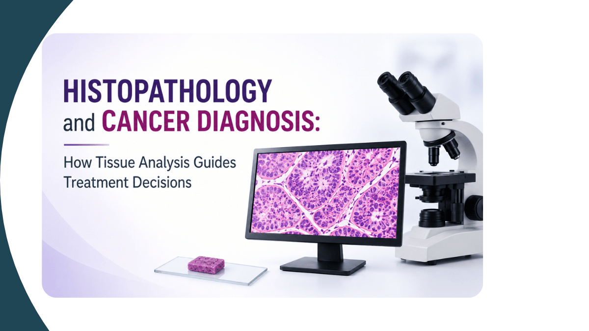
When a patient is suspected of having cancer, the definitive diagnosis almost always begins with tissue. Histopathology - the microscopic examination of tissue samples — is the gold standard for cancer diagnosis. It identifies the type of cancer, its grade and stage, the status of surgical margins, and the molecular characteristics that guide treatment decisions. As a pathologist at PESIMSR, Kuppam, this work is at the centre of what I do every day.
What is histopathology and how does it work?
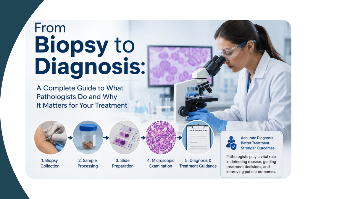
Histopathology is the branch of pathology concerned with the microscopic examination of tissue obtained from biopsy or surgical resection. When a surgeon removes a tumour or takes a tissue sample, that specimen is sent to the pathology laboratory where it undergoes a structured process: grossing (macroscopic examination and sampling), tissue processing, embedding in paraffin wax, sectioning into thin slices, staining — most commonly with haematoxylin and eosin — and microscopic examination by the pathologist.
Each step in this process requires precision and quality control. The quality of tissue processing, staining, and slide preparation directly affects the accuracy of the diagnosis. This is why laboratory quality standards in histopathology are not administrative formalities — they are patient safety requirements.
How does histopathology diagnose cancer?
Cancer diagnosis through histopathology begins with identifying abnormal cells in the tissue sample. The pathologist examines the cellular architecture, nuclear characteristics, mitotic activity, and the relationship between tumour cells and surrounding normal tissue. These features collectively determine whether the tissue is benign, malignant, or of borderline behaviour.
For each tumour, the pathologist reports the histological type — what kind of cell the cancer arose from — and the grade, which reflects how abnormal the cells appear and how aggressively the tumour is likely to behave. In breast cancer, for example, grading using the Nottingham Grading System considers tubule formation, nuclear pleomorphism, and mitotic count. This grade is not just descriptive — it directly influences cancer treatment decisions including chemotherapy protocols and radiation planning.
What types of cancer are diagnosed through histopathology?
Histopathology is used in the diagnosis of virtually all solid tumour types. In my practice the cancers I evaluate most frequently include:
- Breast cancer:- including invasive ductal carcinoma, invasive lobular carcinoma, special type tumours, and in situ lesions. Assessment includes grade, lymphovascular invasion, and receptor status through immunohistochemistry.
- Gynaecological cancers:- cervical carcinoma, endometrial carcinoma, ovarian tumours, and vulvar lesions. Cervical biopsies are evaluated using established classification systems to determine the degree of dysplasia or invasion.
- Skin and dermatopathology:- melanoma, basal cell carcinoma, squamous cell carcinoma, and rare adnexal tumours. Accurate margin assessment in skin excisions is critical for determining whether re-excision is needed.
- Renal tumours:- clear cell carcinoma, papillary renal cell carcinoma, oncocytoma, and nephroblastoma in children. Distinguishing tumour type and grade has direct implications for surveillance and treatment.
- Soft tissue tumours:- a particularly challenging area given the wide range of tumour types, many of which are rare. Immunohistochemistry and molecular markers play an important role in classification.
- Lymph node pathology:- assessment of lymph nodes for metastatic disease, lymphoma, and reactive conditions.
My research interests span several of these areas, including breast cancer histopathology — with published work on tumour budding in invasive breast carcinoma, post-chemotherapy histopathological findings, ABO blood group associations, and cytological grading of proliferative breast lesions.
What is immunohistochemistry and why does it matter in cancer diagnosis?
Standard haematoxylin and eosin staining identifies the morphology of tumour cells. Immunohistochemistry (IHC) goes further — it uses antibodies to detect specific proteins expressed by tumour cells, allowing the pathologist to determine the molecular subtype of the cancer and identify therapeutic targets.
In breast cancer, IHC is used to assess oestrogen receptor (ER), progesterone receptor (PR), and HER2 expression. These results determine whether a patient is eligible for hormone therapy, HER2-targeted therapy, or requires chemotherapy alone. In colorectal cancer, mismatch repair protein testing by IHC identifies microsatellite instability — relevant for immunotherapy eligibility. In poorly differentiated tumours where the cell of origin is unclear, IHC panels help identify the primary site and guide oncology decisions.
The accuracy of IHC results depends on tissue fixation, processing, antibody quality, and interpretation — all areas governed by laboratory quality standards and accreditation requirements.
What does a histopathology report contain and how should it be interpreted?
A histopathology report is a structured document that communicates the pathologist's findings to the clinical team. For a cancer specimen, the report typically includes:
- Macroscopic description - the size, appearance, and sampling of the specimen
- Histological type - the classification of the tumour
- Histological grade - the degree of differentiation
- Margins - whether the tumour has been completely excised or extends to the resection margins
- Lymphovascular invasion - whether tumour cells have entered blood vessels or lymphatics
- Lymph node status - the number of nodes examined and the number containing metastatic tumour
- Immunohistochemistry results - receptor status and other molecular markers where applicable
- Pathological stage - assigned in accordance with TNM staging criteria
Each element of the report contributes to the multidisciplinary team's treatment planning. A complete and accurate pathology report in cancer care is not a formality — it is the document on which surgery, chemotherapy, radiation, and targeted therapy decisions are based.
What should patients know about their biopsy and histopathology report?
Patients often receive their histopathology report before they have had the opportunity to discuss it fully with their clinician. Here are the most important things to understand:
A report takes time for a reason
Tissue processing, embedding, sectioning, staining, and microscopic examination cannot be rushed without compromising accuracy. Standard turnaround times for most biopsies are three to five working days. Complex cases requiring additional stains or immunohistochemistry may take longer. The time taken reflects the thoroughness of the process, not a problem with the sample.
The diagnosis requires clinical correlation
A histopathology report describes what the pathologist sees in the tissue. The final clinical diagnosis integrates the pathology findings with the patient's clinical history, imaging results, and laboratory data. The treating clinician correlates all of this information before making treatment decisions. If the report appears at odds with clinical findings, communication between the clinician and pathologist is essential.
Questions worth asking your doctor about your report
- What is the type of cancer and what does this mean for treatment?
- What is the grade and what does this tell us about how the cancer is likely to behave?
- Are the margins clear? If not, is further surgery needed?
- What do the immunohistochemistry results mean for my treatment options?
- Is a second pathology opinion needed for this case?
Frequently asked questions about histopathology and cancer
Can a biopsy spread cancer?
No. A properly performed biopsy does not spread cancer. This is a common concern but is not supported by evidence. The biopsy is essential for diagnosis — without it, appropriate treatment cannot be planned.
What is the difference between a biopsy and a cytology sample?
A biopsy removes a piece of tissue that is processed and examined as a section. Cytology examines individual cells — from a fine needle aspiration, a Pap smear, or a fluid specimen. Histopathology provides more architectural detail and is generally required for definitive cancer diagnosis and staging. Cytology is faster and less invasive but may not always provide sufficient information for a complete diagnosis.
Is a second opinion on a histopathology report appropriate?
Yes, and in complex or rare cases it is encouraged. Tumour pathology can be challenging, particularly for rare entities or borderline lesions. A second review by an expert pathologist at a specialist centre is a reasonable and appropriate step for patients with complex diagnoses. Many major cancer centres routinely review outside pathology before initiating treatment.
Written by Dr Anikode Subramanian Ramaswamy, MBBS, MD Pathology, ISO 15189 Assessor, Professor and Head of the Department of Pathology, PESIMSR, Kuppam, Andhra Pradesh.
To reach Dr Anikode Subramanian Ramaswamy at PESIMSR, Kuppam.


Add a Comment