Background:
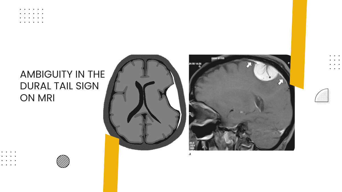
Meningiomas give rise to the dural tail sign (DTS) on contrast-enhanced magnetic resonance imaging (CEMRI). The presence of DTS does not always qualify for a meningioma, as it is seen in only 60-72% of cases. This sign has been described in various other lesions like lymphomas, metastasis, hemangiopericytomas, schwannomas and very rarely glioblastoma multiforme (GBM). The characteristics of dural-based GBMs are discussed here, as only eleven such cases are reported in the literature to date. Here we discuss the unique features of this rare presentation.
Case Description:
A 17-year-old male presented to the emergency department (ED) with complaints of headache, recurrent vomiting, vision loss in the right eye and altered sensorium. On examination, the patient was drowsy with right hemiparesis, secondary optic atrophy in the right eye and papilledema in the left eye. MRI brain showed a heterogeneous, predominantly solid cystic lesion with a central hypo-intense core suggestive of necrosis with heterogeneous enhancement and a positive DTS. Patient underwent emergency left parasagittal parieto-occipital craniotomy and gross total tumour excision, including the involved dura and the falx. On opening the dura, the tumour was surfacing, invading the superior sagittal sinus and the falx, greyish, soft to firm in consistency with central necrosis and highly vascular, suggesting a high-grade lesion. Postoperative computed tomography (CT) of the brain showed evidence of gross total tumour (GTR) excision. The postoperative course of the patient was uneventful. Histopathological analysis revealed GBM with PNET-like components. The dura as well as the falx were involved by the tumour.
Conclusion:
GBMs can arise in typical locations along with DTS mimicking meningiomas. Excision of the involved dura and the falx becomes important in this scenario, so as to achieve GTR. Hence high index of suspicion preoperatively aided by Magnetic Resonance Imaging (MRS) can help distinguish GBMs from meningioma, thereby impacting upon the prognosis.
Keywords: Dural tail sign, glioblastoma multiforme, meningioma, posterior third parasagittal
Refer the link to know more: https://pmc.ncbi.nlm.nih.gov/articles/PMC5875113/
Add a Comment