The vascular system, the network of arteries, veins, and lymphatic vessels that carries blood and fluid throughout the body, is responsible for delivering oxygen and nutrients to every tissue and returning deoxygenated blood and metabolic waste to the lungs and kidneys. When something goes wrong in this system, the consequences range from visible veins on the surface of the legs to life-threatening events such as aortic rupture, pulmonary embolism, or ischaemic stroke. Most vascular conditions that are caught early and managed appropriately do not progress to these extremes. Most that are ignored or attributed to ageing, tiredness, or minor musculoskeletal problems eventually do. As a Consultant Vascular and Endovascular Surgeon at Sparsh Hospital, Hennur, Bengaluru, with DrNB in Vascular and Endovascular Surgery and over 16 years of clinical experience, I see patients every week who have been living with vascular symptoms for months or years before realising that what they were experiencing was a treatable vascular condition rather than an inevitable part of getting older. This guide is for every patient in Bengaluru who has leg symptoms, wound problems, or circulation concerns and wants to understand what those symptoms might indicate and when to seek specialist assessment.
Two Systems, Two Different Problems: Arterial and Venous Disease
The legs have two distinct vascular systems, and understanding the difference between them is the first step in understanding vascular symptoms. The arterial system delivers oxygenated blood from the heart to the tissues under pressure. When arteries become narrowed by atherosclerosis (plaque buildup), blood flow to the tissues is reduced and, if severe enough, the tissue begins to suffer from insufficient oxygen supply. The venous system returns deoxygenated blood from the tissues back to the heart, against gravity, using valves in the vein walls to prevent backflow. When these valves fail, blood pools in the lower leg, pressure builds in the veins, and the consequences range from varicose veins and swelling to skin changes and leg ulcers. Arterial and venous disease can coexist in the same patient, and distinguishing which system is primarily responsible for a patient's symptoms is the essential clinical task at the first vascular consultation.
Varicose Veins: When Cosmetic Concern Becomes a Vascular Problem
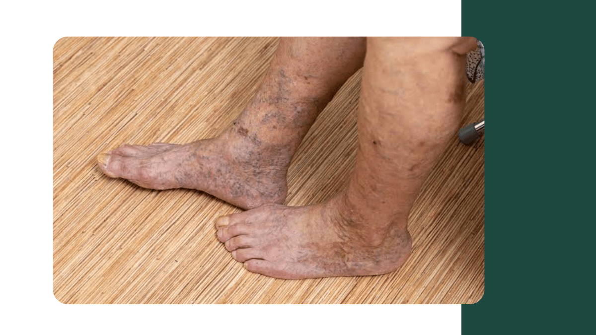
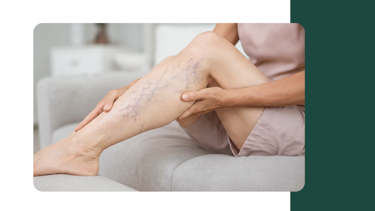
Varicose veins are the most visible vascular condition, and the most commonly dismissed. The enlarged, twisted veins that appear beneath the skin of the legs are not simply a cosmetic problem in the majority of patients who have them. They are the surface expression of venous valve failure that is producing abnormally high pressure throughout the superficial venous system of the leg. This elevated venous pressure is what produces the heaviness, aching, swelling, itching, and night cramps that varicose vein patients describe. It is also what, over years, damages the skin of the lower leg. The skin changes that follow chronic venous hypertension, including thickening, discolouration, eczema-like changes, and eventually ulceration, are not reversible once they are established. Treating the varicose veins before these changes develop is the correct approach, not waiting until the skin has already been permanently damaged.
Modern varicose vein treatment at Sparsh Hospital, Hennur, is minimally invasive and performed under local anaesthesia for the majority of patients, with no hospital stay required. Endovenous laser ablation (EVLA) and radiofrequency ablation (RFA) treat the diseased trunk vein from inside using heat energy delivered through a fine catheter, causing the vein to close permanently without the large incision of traditional stripping surgery. Sclerotherapy treats smaller branch varicosities and reticular veins by injecting a solution that causes the vein to shrink and disappear. A duplex ultrasound scan of the venous system is always the first investigation, because it maps the pattern of reflux, identifies the incompetent valves, and guides the choice of which veins to treat and in what sequence.
Peripheral Arterial Disease: Why Leg Pain on Walking Is Never Just About Age
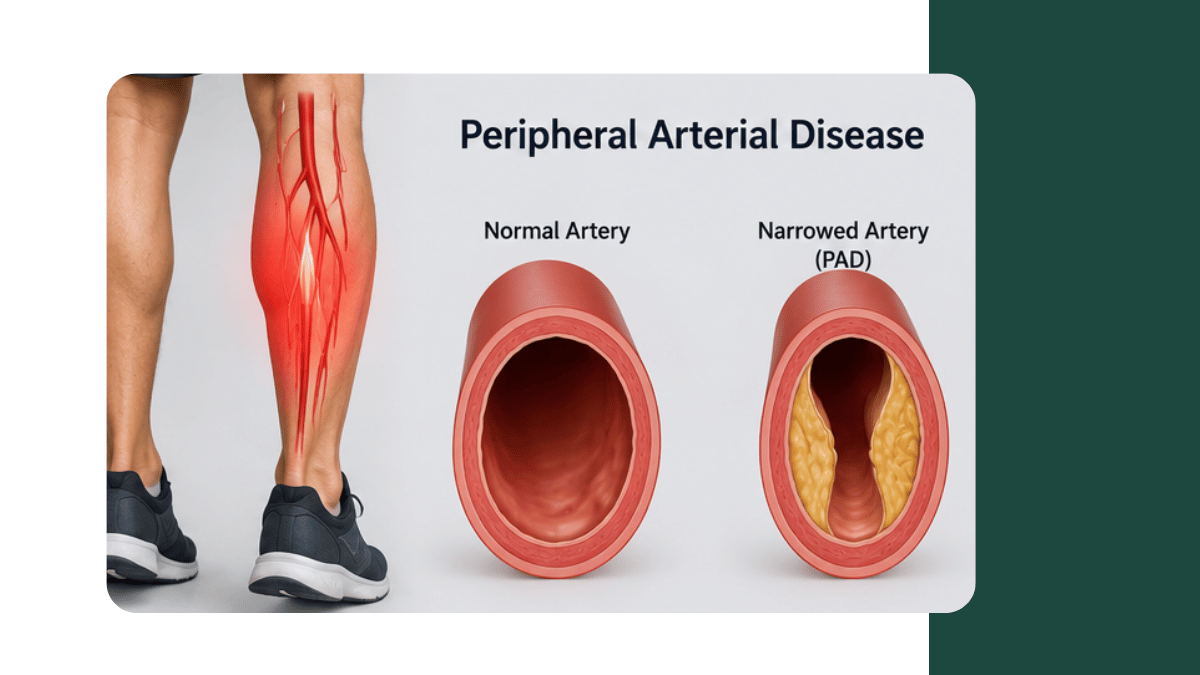
Peripheral arterial disease (PAD) is caused by atherosclerosis of the arteries that supply the legs, progressively narrowing the lumen and reducing blood flow. The cardinal symptom of PAD is intermittent claudication: cramping pain or heaviness in the calf, thigh, or buttock that comes on during walking at a predictable distance and resolves within a few minutes of rest. This pattern is characteristic because, at rest, the reduced blood flow is sufficient for the resting metabolic demand of the muscle. During exercise, when demand increases, the supply cannot keep pace and the muscle cramps. Many patients attribute this symptom to arthritis, age, or fitness and reduce their walking rather than seeking assessment. This is clinically significant because PAD is a marker of systemic atherosclerosis: the patient who has blocked leg arteries very frequently also has atherosclerosis in the coronary arteries and carotid arteries, and their cardiovascular risk management needs to be addressed alongside the peripheral circulation.
The most accessible investigation for PAD is the Ankle Brachial Pressure Index (ABPI), a simple test comparing blood pressure at the ankle and at the arm that provides an objective measure of the degree of arterial obstruction. An ABPI below 0.9 indicates PAD. An ABPI below 0.5 indicates severe PAD with a risk of critical limb ischaemia, where the blood flow is insufficient to maintain tissue viability at rest. Critical limb ischaemia presents as rest pain (pain in the foot at night or at rest that is relieved by hanging the leg down), tissue loss, or gangrene, and represents a vascular emergency requiring urgent revascularisation to save the limb. For PAD patients in Bengaluru, early diagnosis and the appropriate intervention, whether angioplasty, stenting, endarterectomy, or bypass surgery, prevents progression to critical limb ischaemia and dramatically reduces the risk of amputation.
Diabetic Foot and Limb Salvage: The Vascular Dimension of Diabetes
Diabetes damages the blood supply to the feet through two mechanisms that together create a devastating vulnerability. Peripheral arterial disease progresses faster in diabetics and tends to affect the smaller arteries below the knee that supply the foot, making revascularisation technically more demanding. Peripheral neuropathy removes the pain signal that would normally alert a person to a foot injury, blister, or pressure ulcer before it becomes a wound. Together, these mean that foot wounds in diabetics frequently go unnoticed until they are already deep, infected, or gangrenous. India has the world's second largest diabetic population, and diabetic foot complications are the most common cause of non-traumatic lower limb amputation in the country. The vascular surgeon's role in diabetic foot is to assess the blood supply, revascularise the foot where the circulation is insufficient to support healing, and work as part of the multidisciplinary team, alongside the diabetologist, wound care nurse, orthotist, and infectious disease specialist, to save as much of the foot as possible. The outcome of diabetic foot disease is dramatically better when vascular assessment and intervention are integrated from the beginning rather than added late.
Deep Vein Thrombosis: Why Leg Swelling With Calf Pain Is Always Urgent
Deep vein thrombosis (DVT) is the formation of a blood clot within the deep venous system of the leg, most commonly in the calf or thigh. It typically presents as unilateral leg swelling, warmth, and tenderness, often in the calf. DVT is not merely a problem in the leg: the principal danger is pulmonary embolism (PE), which occurs when part of the clot breaks off and travels to the pulmonary arteries in the lungs. Massive PE is immediately life-threatening. Even small PE can cause serious respiratory compromise. DVT is therefore a clinical emergency, not a condition to monitor.
DVT is diagnosed by duplex ultrasound, which is the first investigation of choice in any patient presenting with unilateral leg swelling. Treatment is anticoagulation (blood thinning medication) to prevent clot extension and embolisation, and in selected patients with extensive proximal DVT that is causing severe symptoms or threatening limb viability, catheter-directed thrombolysis (CDT) can be used to actively dissolve the clot through a catheter placed directly in the affected vein. At Sparsh Hospital, Hennur, Bengaluru, I have extensive experience in endovascular management of DVT, including CDT for selected patients with iliofemoral DVT where rapid clot removal reduces the risk of the long-term post-thrombotic syndrome, a chronic condition of venous obstruction and valve damage that causes permanent swelling, pain, and skin changes in the affected leg.
Endovascular Surgery: The Minimally Invasive Revolution in Vascular Care
Endovascular surgery has transformed vascular medicine over the past two decades. Rather than making a large incision to access and repair a blood vessel directly (open surgery), endovascular procedures are performed through small puncture sites using thin catheters, guidewires, and image guidance (fluoroscopy) to navigate inside the blood vessel and deliver treatment to the diseased segment from the inside. The advantages for appropriately selected patients are significant: smaller incisions, less pain, faster recovery, shorter hospital stay, and the ability to treat patients who are not suitable for major open surgery due to medical comorbidities.
The range of conditions treatable by endovascular technique has expanded steadily. Peripheral arterial occlusions are treated by angioplasty (balloon inflation to open the narrowing) and stenting (placing a metallic scaffold to keep the vessel open). Aortic aneurysms, which once required extensive open surgery with a large abdominal or thoracic incision, can in many cases now be treated by EVAR (Endovascular Aneurysm Repair), in which a stent-graft is delivered through the femoral arteries and deployed across the aneurysm to exclude it from the circulation. DVT is treated by catheter-directed thrombolysis. Arteriovenous fistulas that have narrowed and are no longer functioning adequately for haemodialysis are restored by fistula angioplasty. Carotid artery stenosis causing stroke risk is treated by carotid artery stenting as an alternative to open endarterectomy in selected patients. The decision between endovascular and open surgical treatment is not a hierarchy, with endovascular always preferred. It is a clinical decision based on the anatomy of the condition, the patient's overall health, and which approach will deliver the most durable outcome.
Dialysis Access: AV Fistula and the Vascular Surgeon's Role in Kidney Care
Patients with end-stage kidney disease who require haemodialysis need a reliable, durable vascular access point through which large volumes of blood can be circulated through the dialysis machine three times a week. The arteriovenous (AV) fistula, created by surgically connecting an artery and vein in the forearm or upper arm, is the gold standard access for haemodialysis. It uses the patient's own vessels, has the lowest infection and complication rates, and provides the most durable access. Creating, maintaining, and repairing AV fistulas is a significant and technically demanding part of vascular practice. A fistula that has failed to mature after creation, developed a stenosis that reduces flow, clotted, or developed an aneurysm all requires specialist vascular management. Planning the optimal dialysis access requires a vascular surgeon to assess the vessels with duplex ultrasound before creation to select the best site and configuration for that patient's anatomy, and to ensure that every available vessel is preserved for future access as the patient's dialysis journey continues.
When to See a Vascular Surgeon in Bengaluru: Key Symptoms and Warning Signs
The following symptoms in any patient should prompt a vascular surgery consultation rather than a wait-and-see approach.
For venous symptoms: varicose veins associated with aching, swelling, skin changes, or previous bleeding; leg ulcers that have not healed within four weeks; sudden onset unilateral leg swelling with calf tenderness (DVT until proven otherwise, requiring same-day assessment); and chronic swelling of one or both legs affecting quality of life.
For arterial symptoms: calf, thigh, or buttock pain that comes on during walking at a predictable distance and resolves with rest (claudication); foot or calf pain at rest, particularly at night; any colour change in the toes or feet, particularly blue or black discolouration; and cold feet with absent pulses.
For diabetic foot: any foot wound in a diabetic patient that has not improved within two weeks of appropriate wound care; spreading infection, new numbness or tingling, or discolouration around a foot wound.
For all patients: sudden, severe, tearing pain in the chest or abdomen (possible aortic emergency, call emergency services); neck bruit or history of TIA (transient ischaemic attack) requiring carotid artery assessment for stroke prevention; and swelling of the arm or face suggesting superior vena cava obstruction. I consult at Sparsh Hospital Hennur from Monday to Saturday, 9 AM to 4 PM; at Chinmaya Mission Hospital, Indiranagar, on Tuesdays and Fridays from 5 PM; and at Sparsh Hospital Yelahanka on Wednesdays and Saturdays. For patients across north and central Bengaluru, I provide the full spectrum of vascular and endovascular surgical care, from varicose vein treatment and dialysis access to complex limb salvage, aortic surgery, and stroke prevention.
To book a vascular surgery consultation with Dr. Abhilash V at Sparsh Hospital Hennur, Chinmaya Mission Hospital Indiranagar, or Sparsh Hospital Yelahanka, call +91 76766 84611 or +91 88842 70333.
Written by Dr. Abhilash V, MBBS (Kempegowda Institute of Medical Sciences, Rajiv Gandhi University of Health Sciences, Bengaluru, 2010), MS General Surgery (Bangalore Medical College and Research Institute, Rajiv Gandhi University of Health Sciences, Bengaluru, 2015), DrNB Vascular and Endovascular Surgery (National Board of Examinations, New Delhi, 2022), Consultant Vascular and Endovascular Surgeon, Sparsh Hospital Hennur (DivyaSree Avance, Hennur Bagalur Main Road, HBR Layout, Bengaluru 560043, Monday to Saturday, 9:00 AM to 4:00 PM), Chinmaya Mission Hospital (Indiranagar, Bengaluru, Tuesday and Friday, 5:00 PM onwards), and Sparsh Hospital Yelahanka (New Airport Road, Kogilu Cross, Bengaluru 560064, Wednesday and Saturday, 10:00 AM to 1:00 PM and 2:00 PM to 4:00 PM). 16+ years. 10,000+ patients. Phone: +91 76766 84611 / +91 88842 70333.
Related reading
Peripheral Arterial Disease: Why Leg Pain on Walking Is Never Just About Age
Varicose Veins: When Cosmetic Concern Becomes a Vascular Problem

Add a Comment