










chhaya-pathak

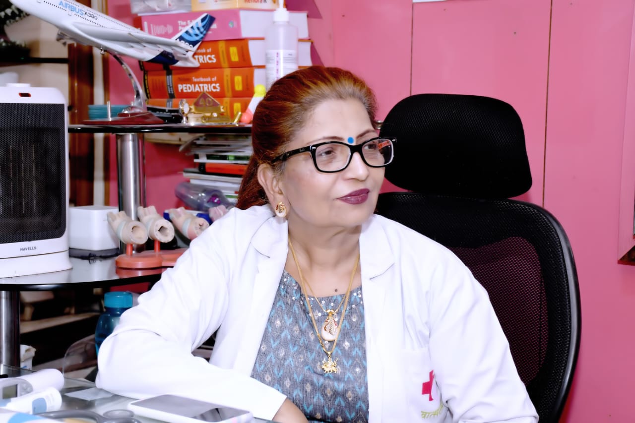
Dr. Chhaya Pathak

Senior Pediatrician, Neonatologist & Intensivist

MBBS, MD (Pediatrics)

40+ Years of Experience

1,00,000+ patients cared

40+ Years of Experience

1,00,000+ patients cared

LinQ Card

About Dr. Chhaya Pathak

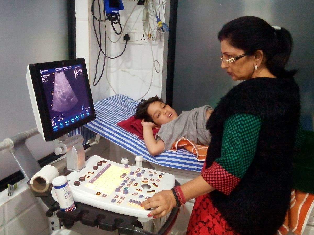
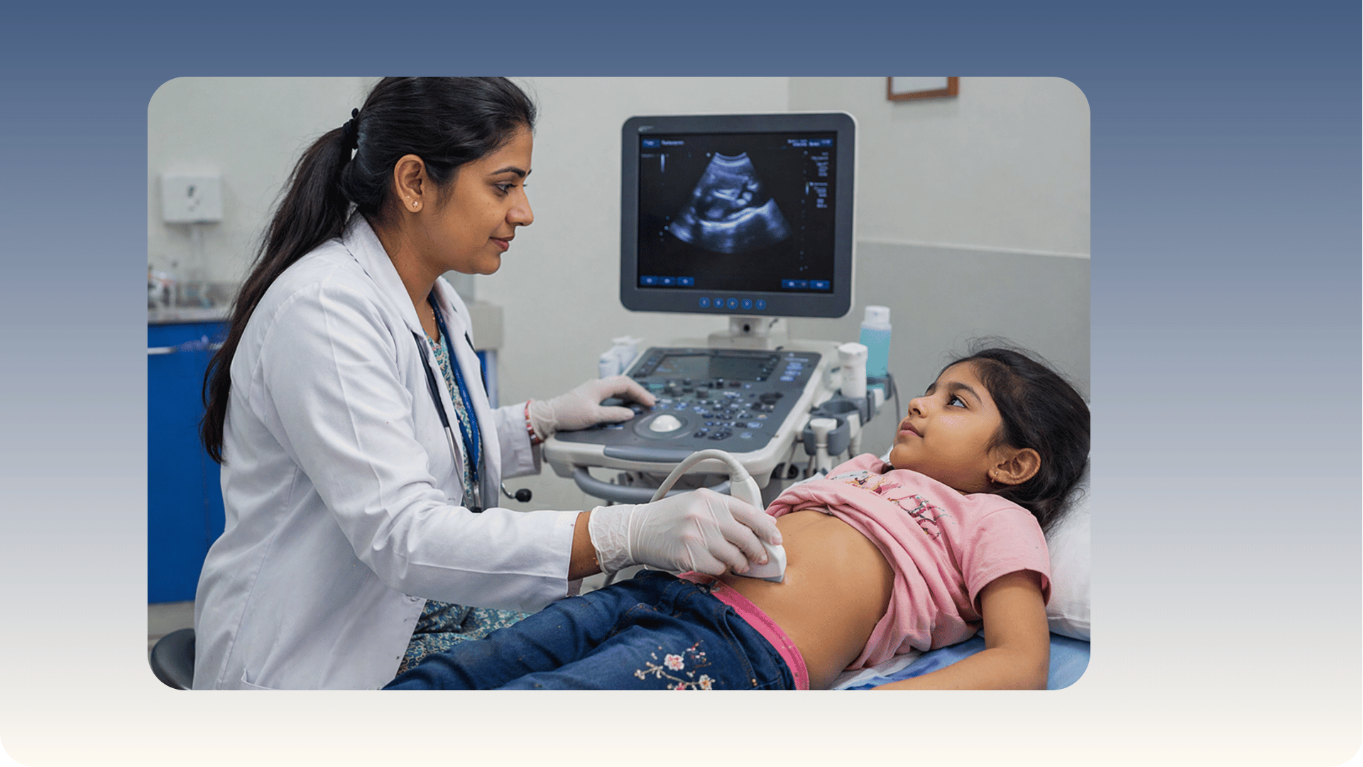
Dr. Chhaya Pathak is a highly accomplished Senior Consultant Pediatrician with over 40 years of clinical excellence, renowned for delivering advanced, compassionate, and evidence-based pediatric care. As the Co-Founder of Vatsalya Children’s Hospital in Varanasi, she has been instrumental in transforming pediatric healthcare in Eastern Uttar Pradesh, introducing cutting-edge technologies such as Point-of-Care Ultrasonography (POCUS) for faster and more accurate bedside diagnosis. Dr. Chhaya Pathak is widely trusted for her expertise in general pediatrics, pediatric critical care, and emergency management, ensuring holistic treatment for infants, children, and adolescents. Her patient-centric approach, combined with decades of experience, has helped thousands of families achieve better child health outcomes. Known for clinical precision and ethical medical practice, Dr. Chhaya Pathak continues to set benchmarks in pediatric healthcare, making her one of the most respected pediatric specialists for comprehensive child care, preventive health, and advanced pediatric treatment solutions.


https://d2vvc091k8tmib.cloudfront.net/s3fs-public/2026-04/Dr.%20Chhaya.jpeg

Dr. Chhaya Pathak specializes in comprehensive pediatric care with a strong focus on pediatric critical care, pediatric emergency management, and advanced bedside diagnostics. With over four decades of experience, Dr. Chhaya Pathak is widely recognized for her expertise in managing complex neonatal and pediatric conditions, ensuring timely intervention and improved clinical outcomes. She is a pioneer in introducing Point-of-Care Ultrasonography (POCUS) in pediatric practice in Eastern Uttar Pradesh, enabling rapid, accurate diagnosis and enhanced decision-making in critical situations. Her specialties also include general pediatrics, growth and developmental monitoring, infectious disease management, and pediatric diagnostic support. Dr. Chhaya Pathak is known for combining clinical excellence with a compassionate, family-centered approach, making her a trusted name for parents seeking reliable and advanced child healthcare solutions. Her commitment to innovation and quality care positions her as a leading pediatric specialist for preventive care, early diagnosis, and comprehensive treatment of childhood illnesses.


Dr. Chhaya Pathak 's Areas of expertise

  • General Pediatrics – Comprehensive diagnosis and treatment of common childhood illnesses with a preventive care approach.
  • Pediatric Critical Care – Expert management of critically ill children requiring intensive monitoring and life-saving interventions.
  • Pediatric Emergency Care – Rapid assessment and treatment of acute pediatric emergencies with precision and efficiency.
  • Point-of-Care Ultrasonography (POCUS) – Advanced bedside imaging for quick, accurate diagnosis and improved clinical decision-making.
  • Neonatal Care – Specialized care for newborns, including management of premature and high-risk infants.
  • Pediatric Infectious Diseases – Diagnosis and treatment of infections with evidence-based protocols for better recovery outcomes.
  • Growth & Development Monitoring – Early detection and management of developmental delays and growth-related concerns.
  • Pediatric Diagnostic Imaging Support – Integration of modern diagnostic tools to enhance accuracy in pediatric evaluations.
  • Preventive Pediatrics & Immunization – Focus on vaccination, nutrition, and preventive strategies to ensure long-term child health.
  • Pediatric Healthcare Program Development – Leadership in establishing and enhancing pediatric care systems and hospital services.
Awards:
  • Recognized as a Pioneer of Pediatric Point-of-Care Ultrasonography (POCUS) in Eastern Uttar Pradesh for advancing bedside diagnostics in pediatric care.
  • Honored for 40+ Years of Clinical Excellence in Pediatrics, reflecting long-standing commitment to child healthcare.
  • Acknowledged for her role as Co-Founder of Vatsalya Children’s Hospital, contributing to the development of one of the region’s leading pediatric healthcare centers.
  • Appreciated for contributions to Pediatric Critical Care and Diagnostic Advancements in the Purvanchal region.
Publications & Academic Contributions:
  • Actively involved in pediatric academic programs and clinical training initiatives.
  • Contributed to educational and training activities associated with pediatric care and ultrasonography (POCUS).
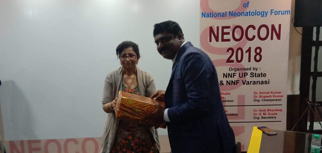
  • Supported National Neonatology Forum (NNF) fellowship and training programs.
  • Participated in knowledge-sharing platforms and continuous medical education (CME) programs in pediatrics.
  • MD (Pediatrics) – Motilal Nehru Medical College, Allahabad
  • MBBS (1982) – Motilal Nehru Medical College, Allahabad

Conditions and Treatments

General Pediatric Care & Common Illness Management

Dr. Chhaya Pathak specializes in managing common pediatric illnesses such as fever, respiratory infections, and gastrointestinal disorders. Her approach focuses on accurate diagnosis and evidence-based treatment for quick recovery. She ensures children receive holistic care with proper nutrition and follow-up support.

Pediatric Emergency & Critical Care Management

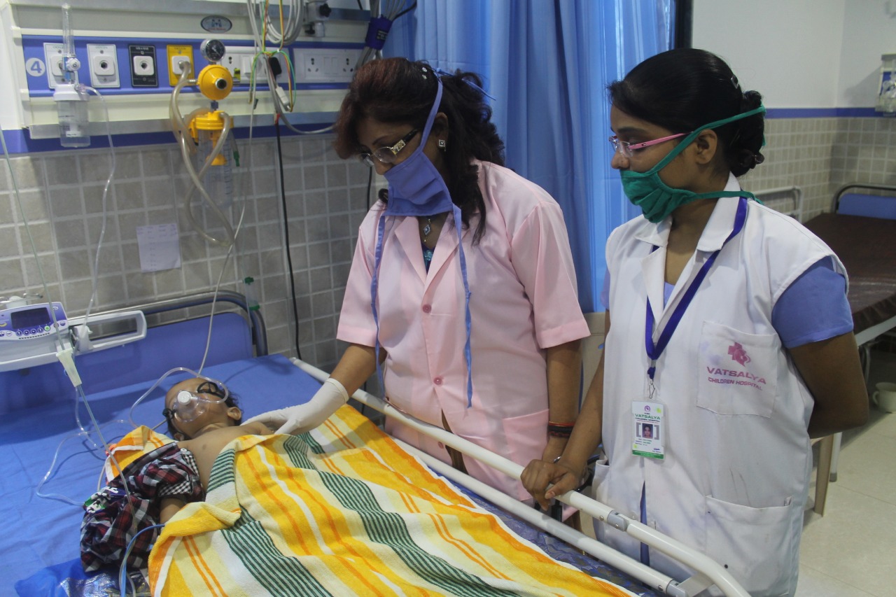
Early diagnosis and timely intervention are key in handling pediatric emergencies and critical conditions. Dr. Chhaya Pathak utilizes advanced clinical expertise and bedside technologies like POCUS for rapid decision-making. Her treatment approach improves survival and recovery outcomes in acute situations.

Neonatal Care & Newborn Health

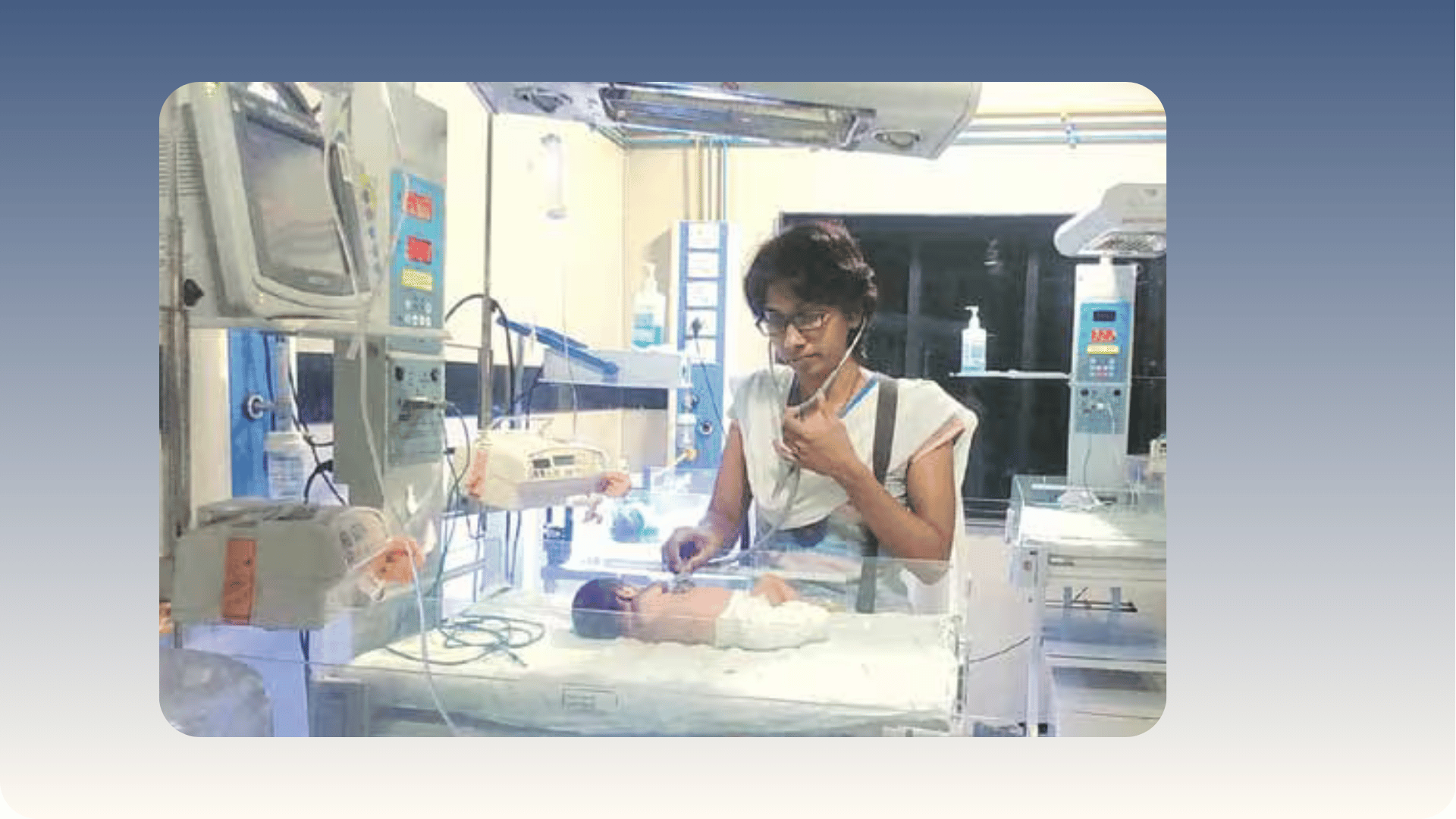
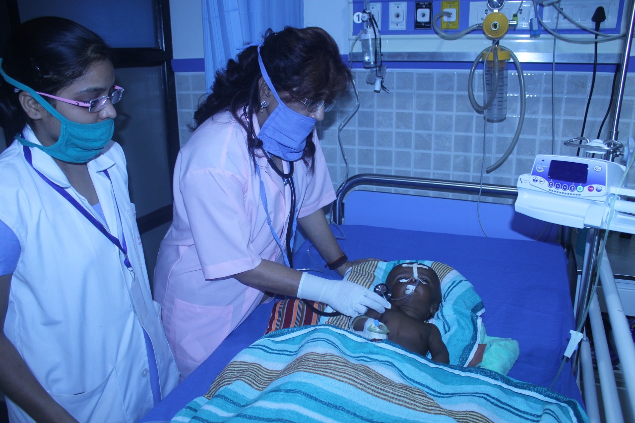
Dr. Chhaya Pathak provides expert care for neonatal conditions including prematurity, low birth weight, and neonatal infections. She emphasizes gentle handling, continuous monitoring, and family-centered care. Her treatment plans are tailored to ensure healthy growth and development from birth.

Pediatric Infectious Disease Management

Managing infectious diseases in children requires precision and preventive strategies. Dr. Chhaya Pathak focuses on accurate identification of infections and targeted therapies to minimize complications. She also guides parents on hygiene and preventive measures for long-term health.

Growth & Development Monitoring

Dr. Chhaya Pathak is experienced in monitoring growth and developmental milestones in children. She identifies delays early and implements corrective interventions through nutrition, therapy, and parental guidance. Her goal is to ensure every child achieves optimal developmental potential.

Preventive Pediatrics & Immunization

Preventive healthcare and immunization are vital for lifelong well-being. Dr. Chhaya Pathak advocates timely vaccinations and routine health check-ups to prevent serious illnesses. She educates families on maintaining a healthy lifestyle and strengthening children’s immunity.

POCUS-Guided Paediatric Emergency Assessment

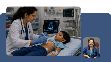
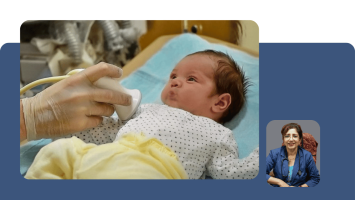
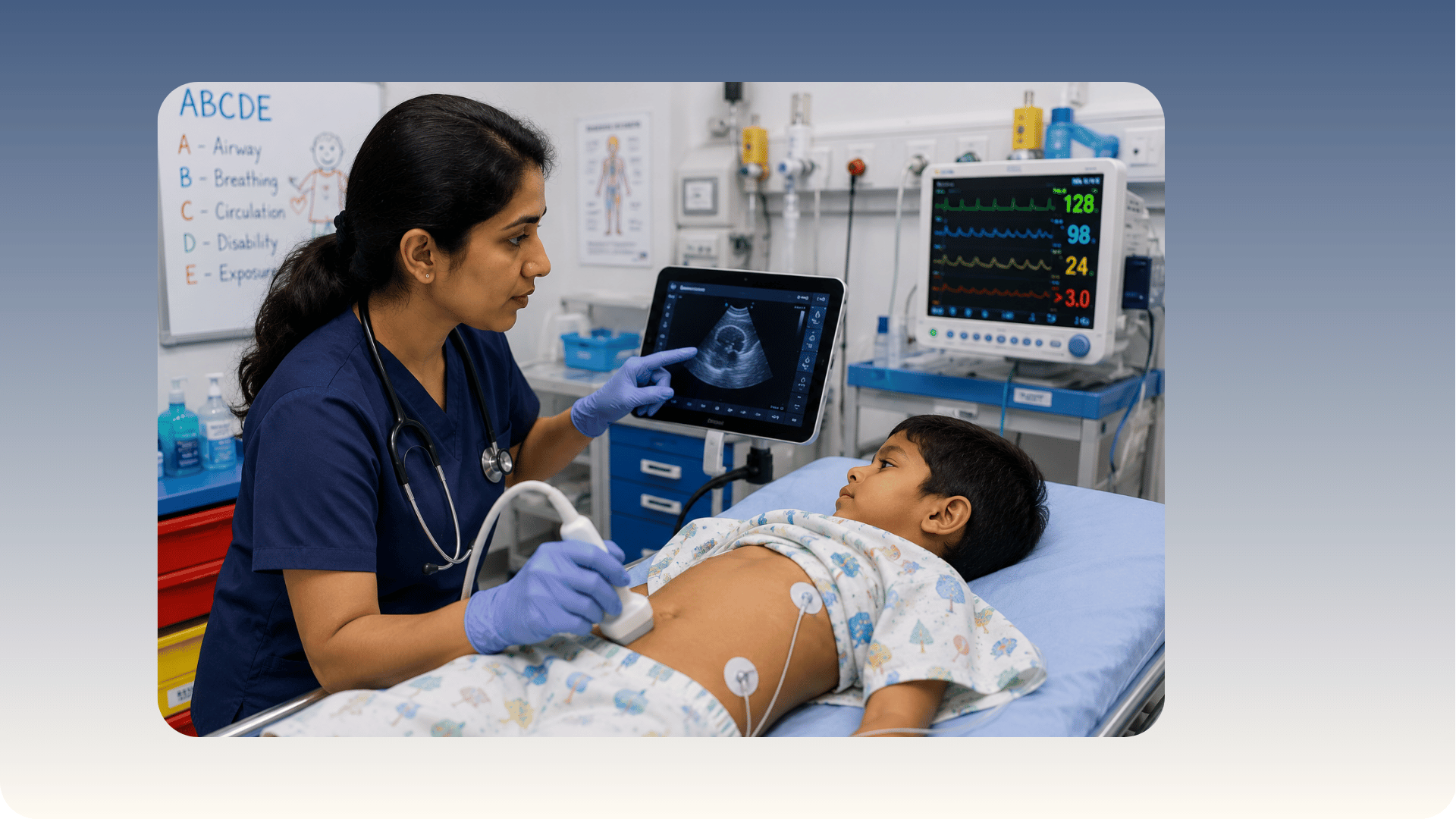
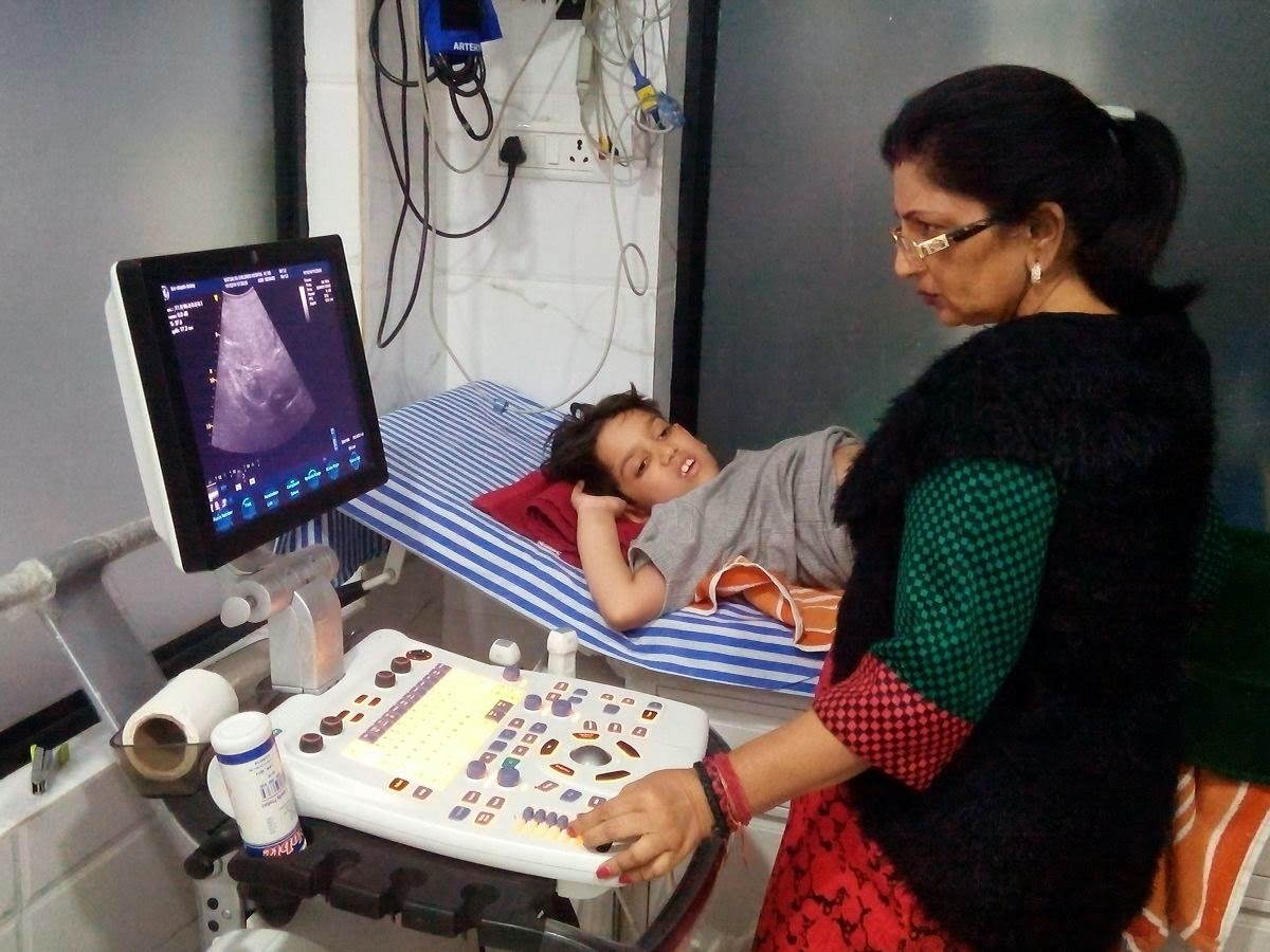
Paediatric emergency assessment guided by Point-of-Care Ultrasonography (POCUS) enables immediate bedside diagnosis of acute respiratory, cardiac, and abdominal conditions in critically ill children without the delay and radiation risk of formal imaging. Dr Chhaya Pathak provides POCUS-guided emergency assessment as a standard part of acute paediatric care at Vatsalya Children's Hospital, Varanasi, using bedside lung, cardiac, and abdominal ultrasound to guide resuscitation, fluid management, and procedural interventions in real time. As the pioneer who introduced paediatric POCUS to clinical practice in Eastern Uttar Pradesh, she brings specific training and extensive clinical experience to every acute paediatric assessment. Written by Dr Chhaya Pathak, MBBS, MD (Pediatrics), Senior Consultant Paediatrician and Co-Founder, Vatsalya Children's Hospital, Mahavir Road, Orderly Bazar, Varanasi, Uttar Pradesh 221002. Pioneer of Paediatric Point-of-Care Ultrasonography (POCUS) in Eastern Uttar Pradesh

Neonatal POCUS and Cranial Ultrasound Assessment

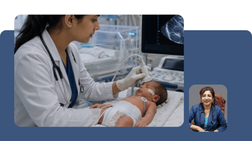
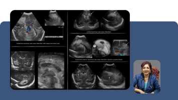
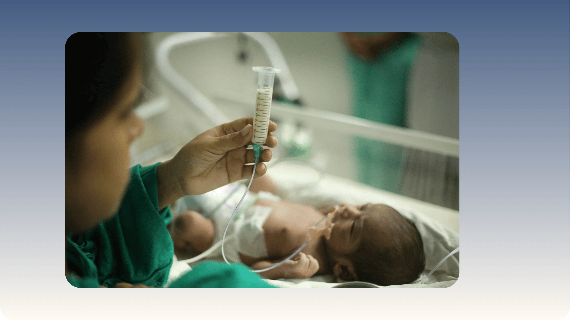
Neonatal POCUS uses the open fontanelle as an acoustic window to assess brain structure and identify intraventricular haemorrhage in preterm infants, while cardiac, lung, and abdominal POCUS guides the management of sick newborns without radiation exposure or the risks of transporting a critically ill neonate to a radiology suite. Dr Chhaya Pathak performs POCUS-guided neonatal assessment at Vatsalya Children's Hospital, Varanasi, including serial cranial ultrasound for preterm infants at risk of intraventricular haemorrhage, cardiac assessment for newborns with suspected congenital heart disease or persistent pulmonary hypertension, and lung assessment for neonates in respiratory distress. This capability is particularly critical in a setting where specialist neonatal radiology is not available around the clock.

POCUS - Point-of-Care Ultrasonography in Paediatric Emergency Care

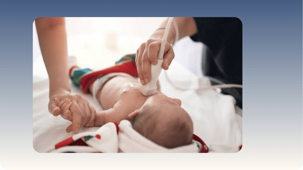
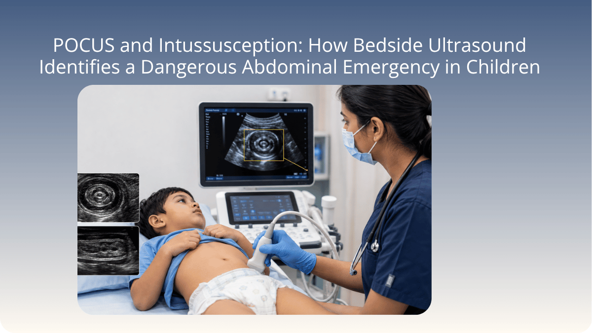
Point-of-Care Ultrasonography - POCUS - is the practice of performing bedside ultrasound at the point of patient contact, in the emergency department, at the bedside, or in the intensive care unit, with results available and interpreted immediately rather than after a formal radiology request and report cycle. In paediatric emergencies, where time from presentation to diagnosis directly affects outcomes, and where the radiation associated with CT scanning is a genuine clinical concern in children, POCUS represents one of the most significant advances in emergency paediatric practice in the past two decades. The clinical value is not only speed - it is the ability to perform a dynamic, real-time examination that answers the specific clinical question being asked without transporting a sick infant from the emergency area. Dr. Chhaya Pathak is the Pioneer of Paediatric POCUS in Eastern Uttar Pradesh, having introduced and developed bedside ultrasonography practice in paediatric emergency and critical care at Vatsalya Children's Hospital, Mahavir Road, Varanasi. With 40 years of paediatric clinical experience and 1,00,000 patients, her clinical judgement determines the POCUS application for each child - and the range of clinical questions POCUS answers in her practice is broad. Intussusception is identified by the target or doughnut sign on abdominal scanning, directing immediate surgical referral and enema reduction without radiology wait. Pleural effusion and pneumonia are assessed with lung POCUS that identifies fluid, consolidation, and B-lines in minutes. Cardiac POCUS assesses pericardial effusion, ventricular function, and volume status in the critically unwell child. Renal POCUS identifies hydronephrosis and urinary tract pathology. And procedural POCUS guides vascular access, diagnostic taps, and drainage procedures with real-time imaging that reduces complication rates. In the context of Eastern Uttar Pradesh, where families frequently travel two to three hours to reach Vatsalya Children's Hospital from district villages, the ability to perform immediate bedside diagnostic imaging at the point of first contact - without radiation, without waiting, and without moving a sick child - is not only clinically superior. It changes outcomes for children whose families could not have made a second journey for a formal radiology appointment at a different facility.

Intussusception and Paediatric Abdominal Emergencies

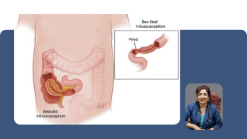
Intussusception - a condition in which one segment of the bowel telescopes into the adjacent segment, progressively compromising its blood supply - is the most common abdominal surgical emergency in children between 6 months and 3 years of age. It is also one of the most consequential conditions to miss, because the window for non-surgical treatment is time-limited and the consequences of bowel necrosis from untreated intussusception are severe. The challenge is that the early presentation of intussusception is easily mistaken for other conditions: an infant who draws up the knees, cries inconsolably for a few minutes, and then appears completely well between episodes may not initially appear to be in an emergency. Dr. Chhaya Pathak, MBBS, MD Paediatrics, 40 years and 1,00,000 patients, and Pioneer of Paediatric POCUS in Eastern Uttar Pradesh, has managed intussusception presentations across four decades of paediatric emergency practice at Vatsalya Children's Hospital, Varanasi. Her POCUS-enabled approach means that when a child presents with episodic colicky abdominal pain and inconsolable crying in the characteristic pattern of intussusception, bedside abdominal ultrasonography is performed immediately - identifying the target or doughnut sign of telescoped bowel on the first clinical contact, in the emergency department, without waiting for formal radiology. This immediate diagnosis allows prompt referral for therapeutic air or hydrostatic enema - the non-surgical reduction technique that resolves intussusception in the majority of uncomplicated cases when it is performed before bowel viability is compromised. Paediatric abdominal emergencies more broadly - including acute appendicitis, malrotation, volvulus, Meckel's diverticulum, and peritonitis - are assessed with the clinical rigour and emergency POCUS capability that 40 years of paediatric experience at a tertiary children's hospital provides. For families in Varanasi and across Eastern Uttar Pradesh who have travelled significant distances with a child in abdominal distress, the ability to reach a diagnosis and initiate the correct management pathway immediately on arrival at Vatsalya Children's Hospital is clinically critical.

Neonatal Respiratory Distress and Lung POCUS: Diagnosing a Sick Newborn's Breathing Problem at the Cot

Respiratory distress is the most common reason a newborn requires admission to a neonatal unit, and its causes are numerous and clinically overlapping. Transient tachypnoea of the newborn (TTN), caused by delayed clearance of fluid from the lungs after delivery, resolves on its own with supportive oxygen. Respiratory distress syndrome (RDS) in premature infants, caused by surfactant deficiency in immature lungs, requires surfactant therapy administered directly into the trachea. Pneumothorax, where air escapes from the lung into the chest cavity and compresses the lung, requires immediate needle decompression. Pneumonia from bacterial or viral infection requires antibiotic therapy. These conditions can look clinically similar in the first minutes and hours of life, yet their treatments are fundamentally different and in some cases directly opposed: giving surfactant to a baby who actually has a pneumothorax can worsen the outcome. The clinical urgency of getting the right diagnosis immediately is therefore acute. Dr. Chhaya Pathak, MD Paediatrics, Senior Consultant Neonatologist and Intensivist, Pioneer of Paediatric POCUS in Eastern Uttar Pradesh, and Co-Founder of Vatsalya Children's Hospital, Varanasi, uses lung POCUS at the cot side to differentiate between these conditions in real time without radiation and without moving the baby. Lung POCUS in the newborn identifies specific ultrasound patterns that are reliably associated with each clinical entity: the absence of normal lung sliding combined with a lung point identifies pneumothorax. A pattern of dense, coalescent B-lines replacing the normal aerated lung appearance indicates either RDS or fluid accumulation from TTN. Consolidation with air bronchograms, where the small airways within the consolidated lung are visible as bright branching lines, indicates pneumonia. These patterns allow a focused lung POCUS performed at the cot in approximately two minutes to redirect the entire management decision from clinical impression to image-guided certainty.

Neonatal Cranial POCUS and Intraventricular Haemorrhage: Protecting the Premature Brain at the Bedside

The brain of a premature infant is one of the most vulnerable structures in neonatology. The germinal matrix, a highly vascular region of undeveloped brain tissue near the fluid-filled ventricles, contains fragile blood vessels that have not yet acquired the structural support of mature cerebral vasculature. Under haemodynamic stress, from fluctuating blood pressure, rapid changes in blood flow during resuscitation, respiratory instability, or handling, these fragile vessels can rupture. The resulting bleed, called intraventricular haemorrhage (IVH), fills the fluid-filled ventricular spaces of the brain with blood. IVH is graded from Grade I (small germinal matrix bleed without ventricular involvement) through Grade II (blood in the ventricles without ventricular dilatation) to Grade III (blood with ventricular dilatation) to Grade IV (blood extending into the brain tissue itself), and this grading directly determines the severity of the prognosis and the monitoring intensity required. Dr. Chhaya Pathak, MD Paediatrics, Senior Consultant Neonatologist and Intensivist, Pioneer of Paediatric POCUS in Eastern Uttar Pradesh, and Co-Founder of Vatsalya Children's Hospital, Varanasi, performs and interprets cranial POCUS through the anterior fontanelle, the soft spot on the top of the skull that remains open in the newborn weeks and provides a direct acoustic window into the brain without requiring X-ray, CT radiation, or transport. At Vatsalya Children's Hospital, serial cranial ultrasound through the fontanelle is part of the standard care protocol for all premature infants at risk, performed on a designated screening schedule to detect IVH at its earliest stage, grade its severity, and monitor for complications including progressive ventricular dilatation and post-haemorrhagic hydrocephalus over the weeks of the neonatal admission. Early detection allows timely neurosurgical referral for hydrocephalus when indicated, developmental follow-up planning to begin from the neonatal period, and informed conversations with families about what the brain scan findings mean for their child's long-term outlook.

Dr. Chhaya Pathak 's Expert Tips & Health Insights

Latest

Articles by Dr. Chhaya Pathak

https://d2vvc091k8tmib.cloudfront.net/s3fs-public/2026-09/Neonatal%20POCUS%20How%20Bedside%20Ultrasound%20Helps%20Diagnose%20and%20Monitor%20the%20Sickest%20Newborns.png
https://d2vvc091k8tmib.cloudfront.net/s3fs-public/2026-04/ALL%20DOCTORS%20PROFILE%20PHOTOS%2026%2012%20%2837%29.png
By: Dr. Chhaya Pathak
04 September 2026

Neonatal POCUS: How Bedside Ultrasound Helps Diagnose and Monitor the Sickest Newborns

A Senior Neonatologist and Pioneer of Paediatric POCUS in Eastern Uttar Pradesh explains how bedside ultrasound is used to diagnose and monitor sick newborns including premature infants, at Vatsalya Children's Hospital, Varanasi.
Read More
https://d2vvc091k8tmib.cloudfront.net/s3fs-public/2026-08/POCUS%20and%20Intussusception%20How%20Bedside%20Ultrasound%20Identifies%20a%20Dangerous%20Abdominal%20Emergency%20in%20Children.png
https://d2vvc091k8tmib.cloudfront.net/s3fs-public/2026-04/ALL%20DOCTORS%20PROFILE%20PHOTOS%2026%2012%20%2837%29.png
By: Dr. Chhaya Pathak
05 August 2026

POCUS and Intussusception: How Bedside Ultrasound Identifies a Dangerous Abdominal Emergency in Children

How bedside POCUS identifies intussusception in infants and children in minutes at Vatsalya Children's Hospital - the target sign, why it matters at night, and why waiting for formal radiology can be dangerous.
Read More
https://d2vvc091k8tmib.cloudfront.net/s3fs-public/2026-07/POCUS%20in%20Paediatrics_%20How%20Bedside%20Ultrasound%20Is%20Changing%20%20the%20Way%20Children%20Are%20Diagnosed%20and%20Treated.png
https://d2vvc091k8tmib.cloudfront.net/s3fs-public/2026-04/ALL%20DOCTORS%20PROFILE%20PHOTOS%2026%2012%20%2837%29.png
By: Dr. Chhaya Pathak
02 July 2026

POCUS in Paediatrics: How Bedside Ultrasound Is Changing the Way Children Are Diagnosed and Treated

When a child arrives in the emergency department with difficulty breathing, abdominal pain, or a high fever and altered behaviour, the clinical question is always the same: what is happening inside this child, and how quickly can I know?
Read More
https://d2vvc091k8tmib.cloudfront.net/s3fs-public/2026-07/Importance%20of%20Vaccination%20in%20Children_%20Preventive%20Pediatric%20Care%20for%20Lifelong%20Immunity.png
https://d2vvc091k8tmib.cloudfront.net/s3fs-public/2026-04/ALL%20DOCTORS%20PROFILE%20PHOTOS%2026%2012%20%2837%29.png
By: Dr. Chhaya Pathak
30 March 2026

Importance of Vaccination in Children: Preventive Pediatric Care for Lifelong Immunity

Vaccination protects children from serious diseases. Dr. Chhaya Pathak, Senior Paediatrician in Varanasi, shares a complete guide on childhood immunisation schedules & preventive care.
Read More
https://d2vvc091k8tmib.cloudfront.net/s3fs-public/2026-07/Child%20Growth%20and%20Development_%20Milestones%2C%20%20Warning%20Signs%2C%20and%20When%20to%20Consult%20a%20Pediatrician.png
https://d2vvc091k8tmib.cloudfront.net/s3fs-public/2026-04/ALL%20DOCTORS%20PROFILE%20PHOTOS%2026%2012%20%2837%29.png
By: Dr. Chhaya Pathak
30 March 2026

Child Growth and Development: Milestones, Warning Signs, and When to Consult a Pediatrician

Dr. Chhaya Pathak, Paediatric Specialist at Vatsalya Children Hospital, Varanasi, guides parents on child growth milestones, warning signs & when to consult a paediatrician.
Read More
https://d2vvc091k8tmib.cloudfront.net/s3fs-public/2026-07/Newborn%20Care%20Guide_%20Managing%20Prematurity%2C%20Low%20Birth%20%20Weight%2C%20and%20Neonatal%20Infections%20Effectively.png
https://d2vvc091k8tmib.cloudfront.net/s3fs-public/2026-04/ALL%20DOCTORS%20PROFILE%20PHOTOS%2026%2012%20%2837%29.png
By: Dr. Chhaya Pathak
30 March 2026

Newborn Care Guide: Managing Prematurity, Low Birth Weight, and Neonatal Infections Effectively

Complete newborn care guide covering prematurity, low birth weight & neonatal infections. Trusted advice by Dr. Chhaya Pathak, Paediatric Specialist, Vatsalya Hospital Varanasi.
Read More
https://d2vvc091k8tmib.cloudfront.net/s3fs-public/2026-07/Pediatric%20Critical%20Care_%20Early%20Signs%2C%20Emergency%20%20Management%2C%20and%20Life-Saving%20Treatments%20for%20Children.png
https://d2vvc091k8tmib.cloudfront.net/s3fs-public/2026-04/ALL%20DOCTORS%20PROFILE%20PHOTOS%2026%2012%20%2837%29.png
By: Dr. Chhaya Pathak
30 March 2026

Pediatric Critical Care: Early Signs, Emergency Management, and Life-Saving Treatments for Children

Dr. Chhaya Pathak, Pediatric Critical Care Specialist at Vatsalya Children Hospital, Varanasi, explains early warning signs, PICU care & life-saving emergency treatments for children.
Read More
https://d2vvc091k8tmib.cloudfront.net/s3fs-public/2026-07/Point-of-Care%20Ultrasonography%20%28POCUS%29%20in%20Pediatrics_%20%20Benefits%20for%20Faster%20Diagnosis%20and%20Better%20Outcomes.png
https://d2vvc091k8tmib.cloudfront.net/s3fs-public/2026-04/ALL%20DOCTORS%20PROFILE%20PHOTOS%2026%2012%20%2837%29.png
By: Dr. Chhaya Pathak
30 March 2026

Point of Care Ultrasonography (POCUS) in Pediatrics: Benefits for Faster Diagnosis and Better Outcomes

Aim & Purpose: Point-of-Care Ultrasonography (POCUS) in pediatrics is revolutionizing bedside diagnosis by enabling faster, safer, and more accurate clinical decisions. It plays a vital role in pediatric emergency ultrasound, offering immediate insights without delays.
Read More
https://d2vvc091k8tmib.cloudfront.net/s3fs-public/2026-07/Pediatric%20Emergency%20Care_%20Recognizing%20Early%20Warning%20%20Signs%20and%20When%20to%20Seek%20Immediate%20Treatment%20for%20Children.png
https://d2vvc091k8tmib.cloudfront.net/s3fs-public/2026-04/ALL%20DOCTORS%20PROFILE%20PHOTOS%2026%2012%20%2837%29.png
By: Dr. Chhaya Pathak
30 March 2026

Pediatric Emergency Care: Recognizing Early Warning Signs and When to Seek Immediate Treatment for Children

Breathing difficulty, seizures, bluish lips — know the early warning signs of pediatric emergencies. Trusted expert care by Dr. Chhaya Pathak, Vatsalya Children Hospital, Varanasi.
Read More

Dr. Chhaya Pathak 's Patient Testimonial Reviews

Leave a Review

Patient Reviews

Dr. Chhaya Pathak

Leave a Review

Frequently Asked Questions

Frequently Asked Questions

View all

What is an intraventricular haemorrhage and how is it detected in a premature baby?

Intraventricular haemorrhage, abbreviated as IVH, is a bleed that occurs inside or around the fluid-filled chambers of the brain called ventricles, and it is one of the most significant complications of premature birth. To understand why premature babies are at risk, it helps to understand what makes the premature brain structurally different from the brain of a full-term newborn. In the premature infant, particularly those born before 32 weeks of gestation, there is a region of the developing brain near the ventricles called the germinal matrix, which contains an extremely rich network of fragile, thin-walled blood vessels. These vessels are actively involved in the development of the brain, but they have not yet acquired the structural reinforcement that mature cerebral blood vessels have. When a premature baby experiences haemodynamic stress, fluctuating blood pressure, a rapid change in blood oxygen level, or the physiological disturbance of a resuscitation, these fragile vessels can rupture. The blood that escapes flows into the ventricular spaces, producing an IVH. IVH is graded by severity: Grade I is a small bleed confined to the germinal matrix itself, without blood entering the ventricles. Grade II is blood entering the ventricles without causing them to dilate. Grade III is blood with ventricular dilatation, which can impair the drainage of cerebrospinal fluid. Grade IV is the most severe, where blood extends directly into the brain tissue. This grading matters because it correlates with long-term neurological outcome: Grade I and II bleeds may have minimal long-term consequences; Grade III and IV carry a higher risk of developmental difficulties, cerebral palsy, or need for neurosurgical intervention for hydrocephalus. Detection is by cranial ultrasound through the anterior fontanelle, the soft open spot on the top of the skull that persists for weeks in premature babies and provides a natural acoustic window into the brain. "At Vatsalya Children's Hospital, Varanasi, serial cranial ultrasound is part of the standard care protocol for every premature baby at risk, and I perform these scans at the cot as part of the routine neonatal assessment, without transport or radiation."

Related reading: 

Neonatal POCUS: How Bedside Ultrasound Helps Diagnose and Monitor the Sickest Newborns
POCUS in Paediatrics: How Bedside Ultrasound Is Changing the Way Children Are Diagnosed and Treated
Point of Care Ultrasonography (POCUS) in Pediatrics: Benefits for Faster Diagnosis and Better Outcomes
Newborn Care Guide: Managing Prematurity, Low Birth Weight, and Neonatal Infections Effectively


Written by Dr. Chhaya Pathak, MBBS, MD (Paediatrics), Senior Consultant Paediatrician, Neonatologist and Intensivist, Pioneer of Paediatric POCUS in Eastern Uttar Pradesh, Co-Founder, Vatsalya Children's Hospital, Varanasi 221002. Phone: +91 9838585111. 

My premature baby is having difficulty breathing. How does a doctor know what is wrong with the lungs?

The question every parent is asking when their premature baby is in respiratory distress is the same question I am asking as the neonatologist: what specifically is causing this breathing problem, and what does it need? A premature baby in respiratory distress is working hard to breathe, with rapid breathing, visible recession of the chest wall with each breath, and often a grunting sound that the baby makes in an attempt to keep the airways open. These signs tell me the baby's lungs are struggling, but they do not tell me why. The answer to why changes the treatment entirely. A premature infant born at 28 to 30 weeks of gestation has lungs that have not yet produced adequate surfactant, the substance that keeps the tiny air sacs from collapsing with every breath. This is respiratory distress syndrome (RDS), and it is treated with exogenous surfactant administered directly into the trachea through a fine tube. A baby with a pneumothorax, where air has escaped from the lung into the chest cavity and is compressing the remaining lung, needs immediate needle decompression of that air. A baby with pneumonia from an infection needs antibiotics. The clinical signs of all three can overlap in the first hours of life. Lung POCUS allows me to distinguish between these entities immediately, at the cot, without moving the baby. The specific ultrasound patterns associated with each diagnosis are distinct: RDS produces a characteristic pattern of dense B-lines replacing the normal air-filled lung appearance. Pneumothorax produces absence of the normal movement of the lung with breathing, combined with a specific finding called the lung point. Pneumonia produces consolidation, where the affected lung segment appears solid rather than aerated. At Vatsalya Children's Hospital, Varanasi, a lung POCUS in my hands at the cot side takes approximately two minutes and gives me the information I need to direct the treatment without delay.

Related reading: 

Neonatal POCUS: How Bedside Ultrasound Helps Diagnose and Monitor the Sickest Newborns
POCUS in Paediatrics: How Bedside Ultrasound Is Changing the Way Children Are Diagnosed and Treated

Point of Care Ultrasonography (POCUS) in Pediatrics: Benefits for Faster Diagnosis and Better Outcomes
Newborn Care Guide: Managing Prematurity, Low Birth Weight, and Neonatal Infections Effectively
Written by Dr. Chhaya Pathak, MBBS, MD (Paediatrics), Senior Consultant Paediatrician, Neonatologist and Intensivist, Pioneer of Paediatric POCUS in Eastern Uttar Pradesh, Co-Founder, Vatsalya Children's Hospital, Varanasi 221002. Phone: +91 9838585111. 

What is the target sign in POCUS and what does it tell the doctor about intussusception?

The target sign - also called the doughnut sign - is the ultrasound finding that identifies intussusception on point-of-care ultrasonography. When one segment of the bowel telescopes into an adjacent segment, as occurs in intussusception, scanning across the affected area in the transverse plane produces a characteristic appearance of concentric rings: the outer wall of the receiving bowel, the mesenteric fat and wall of the intussuscepted segment, and further inner rings from the layers of telescoped bowel. The result on the ultrasound screen is a circular pattern of alternating echogenicities - dark and bright rings - that resembles a target or cross-section of a doughnut. This sign has high sensitivity and specificity for intussusception when it is identified by a trained examiner - in other words, when it is present, it is almost always intussusception, and when intussusception is present, it is almost always visible on POCUS in experienced hands. The clinical importance is that this finding is available immediately, at the bedside, in the emergency department, without the child needing to be moved to a radiology department, without radiation exposure, and without waiting for a formal ultrasound request and report. At Vatsalya Children's Hospital, Varanasi, when I identify the target sign on bedside POCUS in an infant with episodic colicky pain, the management pathway begins at that moment - surgical referral and preparation for therapeutic enema reduction, rather than hours of observation while waiting for formal imaging.

Written by Dr. Chhaya Pathak, MBBS, MD (Pediatrics), Senior Consultant Paediatrician, Neonatologist and Intensivist, Pioneer of Paediatric POCUS in Eastern Uttar Pradesh, Co-Founder, Vatsalya Children's Hospital, Mahavir Road, Orderly Bazar, Varanasi, Uttar Pradesh 221002. 40+ years, 1,00,000+ patients. Phone: +91 9838585111.

Related reading:

POCUS and Intussusception: How Bedside Ultrasound Identifies a Dangerous Abdominal Emergency in Children

POCUS in Paediatrics: How Bedside Ultrasound Is Changing the Way Children Are Diagnosed and Treated

What is the treatment for intussusception and does every child need surgery?

Most children with intussusception diagnosed promptly do not require surgery - this is one of the important things for families to understand, because the word 'surgical emergency' understandably provokes significant alarm. The first-line treatment for uncomplicated intussusception is therapeutic enema reduction - either air enema or hydrostatic (saline or contrast) enema - performed under imaging guidance, typically fluoroscopy or ultrasound. Air or fluid is introduced into the rectum under controlled pressure, and this pressure is transmitted proximally through the bowel, pushing the telescoped segment back to its normal position. The procedure is performed by a paediatric surgeon or radiologist with appropriate facilities, takes approximately 30 to 60 minutes, and in suitable cases successfully reduces intussusception without any incision. The success rate for enema reduction is high in children without signs of bowel compromise - typically 70 to 90 percent in published series. The conditions that predict lower enema success rates and make surgery more likely are: longer duration of symptoms before presentation, evidence of bowel ischaemia or peritonitis, the presence of a lead point (an underlying abnormality that has initiated the intussusception, more common in older children), and failed enema after one or two attempts. Surgery is also required when enema reduction succeeds but the intussusception recurs multiple times. The critical factor that most determines whether enema reduction succeeds is time - the earlier intussusception is diagnosed and referred for enema reduction, the higher the chance of avoiding surgery and the lower the risk of complications.

Written by Dr. Chhaya Pathak, MBBS, MD (Pediatrics), Senior Consultant Paediatrician, Neonatologist and Intensivist, Pioneer of Paediatric POCUS in Eastern Uttar Pradesh, Co-Founder, Vatsalya Children's Hospital, Mahavir Road, Orderly Bazar, Varanasi, Uttar Pradesh 221002. 40+ years, 1,00,000+ patients. Phone: +91 9838585111.

My infant is screaming and drawing up their knees but seems fine in between - could this be intussusception?

The pattern you are describing - an infant who suddenly cries intensely, draws up the knees, and appears to be in severe pain for a few minutes, and then returns to appearing normal until the next episode - is the classic presentation of intussusception, and in a child between 6 months and 3 years of age, this pattern warrants urgent paediatric assessment on the same day. The episodic nature occurs because the cramping pain of the intestinal muscle contracting around the telescoped bowel segment comes in waves, corresponding to peristaltic contractions. Between contractions, the pain is absent and the infant may look completely well - sometimes even smile or feed. This appearance of well-being in the intervals is one of the features that can mislead: the infant does not look sick, and in an environment without immediate investigation capability, the episode may be attributed to colic and the child sent home. What changes as intussusception progresses is the inter-episode interval: the infant becomes more lethargic and less responsive, the normal-interval appearance fades, and pallor becomes more evident. The ominous late sign is the red currant jelly stool - blood-stained mucus passed rectally - which indicates that bowel blood supply is already compromised. At this stage, the window for non-surgical enema reduction has often passed. The most important clinical message is: if your infant has this episodic pattern of intense crying with intervals of normality, seek urgent paediatric assessment immediately - do not wait to see if the episodes resolve on their own. 
Written by
Dr. Chhaya Pathak, MBBS, MD (Pediatrics), Senior Consultant Paediatrician, Neonatologist and Intensivist, Pioneer of Paediatric POCUS in Eastern Uttar Pradesh, Co-Founder, Vatsalya Children's Hospital, Mahavir Road, Orderly Bazar, Varanasi, Uttar Pradesh 221002. 40+ years, 1,00,000+ patients. Phone: +91 9838585111.

Why is POCUS better than X-ray or CT scan for diagnosing intussusception in children?

The comparison between POCUS, X-ray, and CT for intussusception diagnosis in children involves three factors: accuracy, radiation, and time. Plain abdominal X-ray has poor sensitivity for intussusception - it may show features suggestive of bowel obstruction in advanced cases, but in the early presentation, when intervention is most effective, the X-ray is frequently normal or shows only non-specific bowel gas patterns. A normal X-ray does not exclude intussusception. CT scanning of the abdomen does diagnose intussusception accurately, but it delivers a significant radiation dose to a young child's abdomen and pelvis - radiation that carries a long-term cancer risk that is small but real and is clinically unjustifiable when an equally accurate, radiation-free alternative is available. CT also requires the child to be moved from the emergency area, potentially distressing an already unwell infant, and may require sedation. POCUS at the bedside, when performed by an experienced examiner, has sensitivity and specificity for intussusception that is comparable to formal ultrasound. It requires no radiation, no patient transport, no sedation, and no delay. The result is available immediately and the management decision follows within the same assessment. In the context of paediatric emergency care in Varanasi, where the decision to observe versus refer for immediate enema reduction versus prepare for surgery may need to be made within the first hour of a child's arrival, POCUS gives the clinical team the information needed to make that decision without delay. 
 

Written by Dr. Chhaya Pathak, MBBS, MD (Pediatrics), Senior Consultant Paediatrician, Neonatologist and Intensivist, Pioneer of Paediatric POCUS in Eastern Uttar Pradesh, Co-Founder, Vatsalya Children's Hospital, Mahavir Road, Orderly Bazar, Varanasi, Uttar Pradesh 221002. 40+ years, 1,00,000+ patients. Phone: +91 9838585111.

Related reading 

POCUS and Intussusception: How Bedside Ultrasound Identifies a Dangerous Abdominal Emergency in Children

POCUS in Paediatrics: How Bedside Ultrasound Is Changing the Way Children Are Diagnosed and Treated

What is the target sign in POCUS and what does it tell the doctor about intussusception?

The target sign - also called the doughnut sign - is the ultrasound finding that identifies intussusception on point-of-care ultrasonography. When one segment of the bowel telescopes into an adjacent segment, as occurs in intussusception, scanning across the affected area in the transverse plane produces a characteristic appearance of concentric rings: the outer wall of the receiving bowel, the mesenteric fat and wall of the intussuscepted segment, and further inner rings from the layers of telescoped bowel. The result on the ultrasound screen is a circular pattern of alternating echogenicities - dark and bright rings - that resembles a target or cross-section of a doughnut. This sign has high sensitivity and specificity for intussusception when it is identified by a trained examiner - in other words, when it is present, it is almost always intussusception, and when intussusception is present, it is almost always visible on POCUS in experienced hands. The clinical importance is that this finding is available immediately, at the bedside, in the emergency department, without the child needing to be moved to a radiology department, without radiation exposure, and without waiting for a formal ultrasound request and report. At Vatsalya Children's Hospital, Varanasi, when I identify the target sign on bedside POCUS in an infant with episodic colicky pain, the management pathway begins at that moment - surgical referral and preparation for therapeutic enema reduction, rather than hours of observation while waiting for formal imaging.

Written by Dr. Chhaya Pathak, MBBS, MD (Pediatrics), Senior Consultant Paediatrician, Neonatologist and Intensivist, Pioneer of Paediatric POCUS in Eastern Uttar Pradesh, Co-Founder, Vatsalya Children's Hospital, Mahavir Road, Orderly Bazar, Varanasi, Uttar Pradesh 221002. 40+ years, 1,00,000+ patients. Phone: +91 9838585111.

Link copied to clipboard!

Reach Dr. Chhaya Pathak

Vatsalya Children Hospital

Mahavir Road, near Mahavir Mandir Crossing, Orderly Bazar, Varanasi, Uttar Pradesh 221002