
Who is Dr. Murali Mohan S?
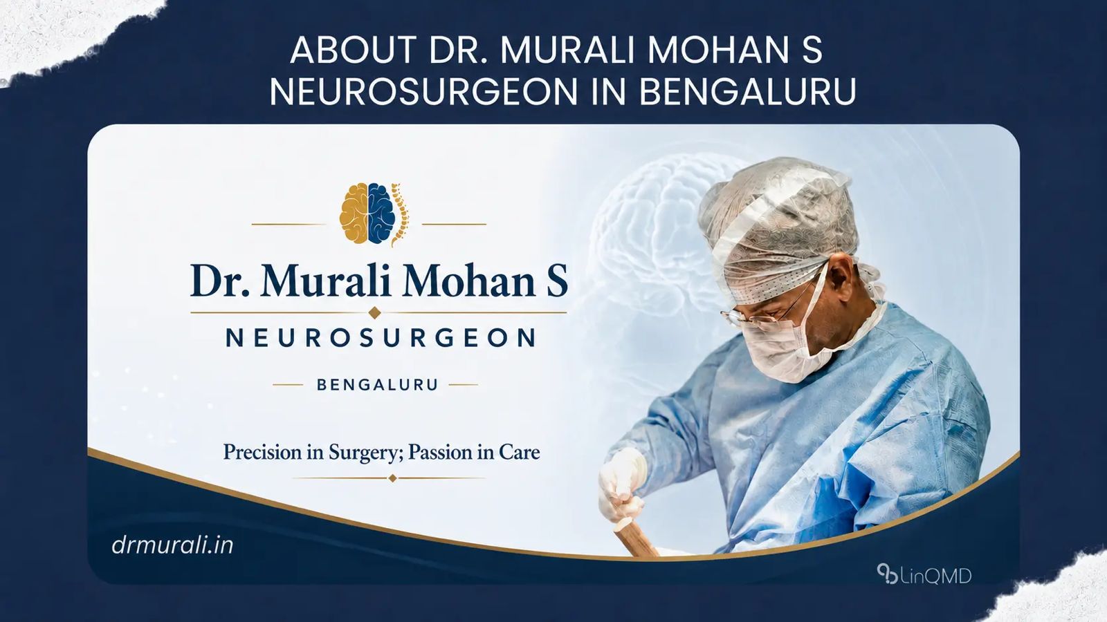
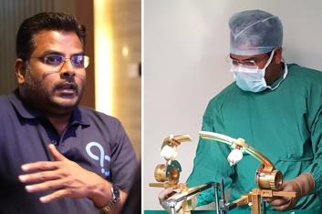
Dr. Murali Mohan S is a consultant neurosurgeon based in Bengaluru. He serves as Lead Consultant, Neurosciences at SPARSH Hospital, Hennur, and also consults at Synapse Clinics in Jayanagar and Yelahanka.
MBBS, DNB (Neurosurgery)
Scan to Connect
LinQ Card
Dr. Murali Mohan S is a leading Neurosurgeon, Healthcare Entrepreneur, Innovator, and an influential thought leader advancing brain and spine care.
Scan to Connect with
Dr. Murali Mohan S
Scan with phone camera


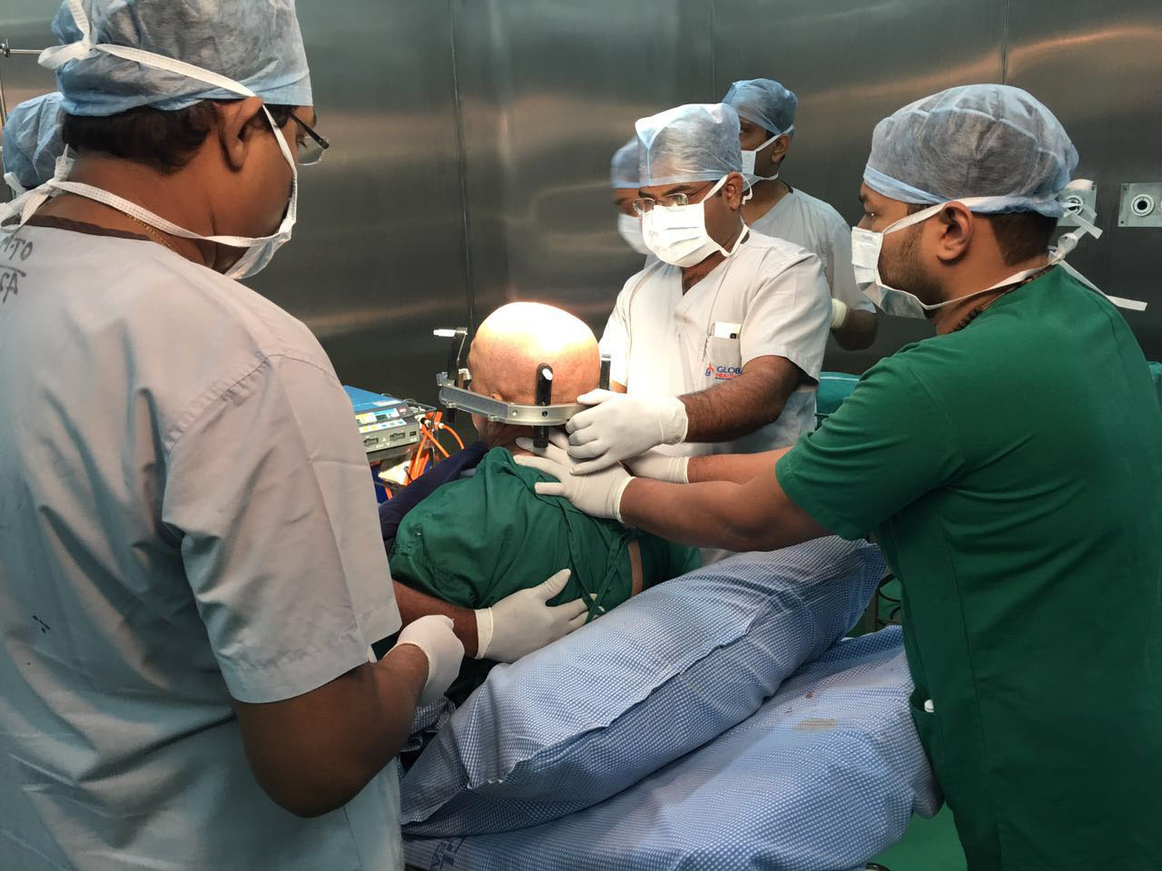
Dr. Murali Mohan S is a leading Neurosurgeon, Healthcare Entrepreneur, Innovator, and an influential thought leader advancing brain and spine care. With over 8,000 brain and spine surgeries performed. He has earned recognition for his surgical expertise in Complex Neurosurgical Procedures. He holds a patent for an indigenous stereotactic frame, founded Dr. Klinisch Research Pvt Ltd. for clinical research, and created LinQMD, an AI-powered platform for doctors’ digital presence. He currently serves as Lead Consultant, Neurosciences at Sparsh Hospital, Hennur. Beyond the operating room, Dr. Murali is a TEDx speaker, blogger, biker, and astronomy enthusiast—leading with ‘clarity, curiosity, and vision across healthcare innovation, research, and communication.
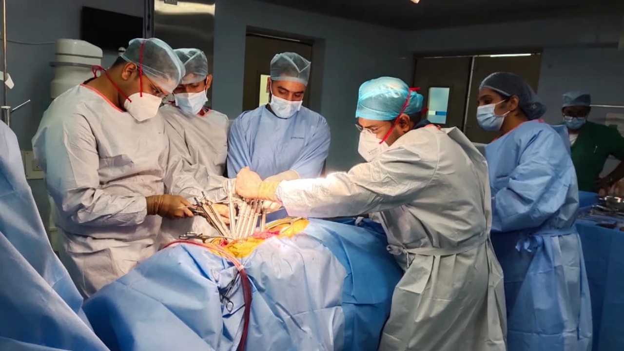
Dr. Murali Mohan S offers comprehensive neurosurgical expertise across brain and spine conditions, combining advanced surgical techniques with patient centered care. His work spans pediatric, adult, and geriatric neurosurgery, with a special focus on complex, minimally invasive, and reconstructive procedures. With over 8,000 surgeries, Dr. Murali is trusted for delivering precision outcomes across a wide spectrum of neurological conditions.
Dr. Murali Mohan S has contributed extensively to neurosurgical research, clinical publications, and academic discourse, with award-winning papers, peer-reviewed journal articles, and textbook chapters. His work reflects a deep commitment to advancing neurosurgical knowledge and practice.
Suboccipital Segment of Vertebral Artery – A Cadaveric Study Neurological Society of India Conference, Madurai (Dec 2006)
Rhino-cerebral Fungal Granuloma, Skull Base Society of India and WFNS Conference, New Delhi (Oct 2007)
Surgery of Craniovertebral Junction, Skull Base Society of India and WFNS Conference, Jaipur (Oct 2016)
Suprasellar Germ Cell Tumor… J Pediatr Endocrinol Metab, 2003
Intracranial Epithelioid Hemangioendothelioma… Childs Nerv Syst, 2008
Pediatric Medulloblastoma: Review of 67 Cases… Asian J Neurosurg, 2008
Suboccipital Segment of Vertebral Artery… Neurol India, 2009
Invasive Rhino-Cerebral Fungal Granuloma… Neurol India, 2010
Morphometric Analysis of Thoracic Pedicle… Neurol India, 2010
Intraoperative Angiography in Cerebral Aneurysm Surgery… Neurol India, 2010
"A Useful Noise in the Operating Room"… J Cerebrovasc Sci, 2015
“Retraction-less Aneurysm Surgery”… J Cerebrovasc Sci, 2015
Pial AV Fistula Presenting as Lobar Hemorrhage… J Cerebrovasc Sci, 2015
Intraventricular Gliosarcomas… World Neurosurg, 2016
Surgical Anatomy of Posterior Third Ventricle – Textbook of Operative Neurosurgery
Empty Sella Syndrome – Ramamurthi and Tandon Textbook of Neurosurgery
Acoustic Neurofibroma – Ramamurthi and Tandon Textbook of Neurosurgery
Stereotaxy for Brain Tumors – Ramamurthi and Tandon Textbook of Neurosurgery
Diencephalic Syndrome – Ramamurthi and Tandon Textbook of Neurosurgery
Bacterial Infections of the Spine – Text of Neurosurgery
Tuberculosis of CNS – Text of Neurosurgery
Information Systems for Knowledge Management… – Knowledge Organisation
Anatomy of the Sellar and Suprasellar Region – Textbook of Endocrinology
Pituitary Adenoma; Growth Hormone Secreting Pituitary Adenomas; Corticotroph Adenomas; Prolactinoma; Other Pituitary Adenomas – Textbook of Endocrinology
Endoscopic Lumbar Discectomy; Endoscopic Surgery for Pituitary Tumors – Clinical Neuroendoscopy
Neurological Society of India
Skull Base Surgery Society of India
Association of Spinal Surgeons of India
Bangalore Neurological Society
Indian Federation of Neuro-endoscopy
Indian Association of Pediatric Neurosurgery
Major Akshay Girish Kumar Rd, Yelahanka Satellite Town, Yelahanka, Bengaluru, Karnataka 560064
Phone: +91 80501 57007 | +91 99007 01080
Email: [email protected]
771, 10th Main Road, 34th Cross Rd, 4th Block, Jayanagar, Bengaluru, Karnataka 560011
Phone: +91 99027 97944 | +91 99007 01080
Email: [email protected]
HBR Layout, Hennur Road, Bengaluru - 560 043
Phone: +91 9483861505
Email: [email protected]
5 reviews • 5★

a month ago

a month ago

a month ago

a month ago

5 months ago
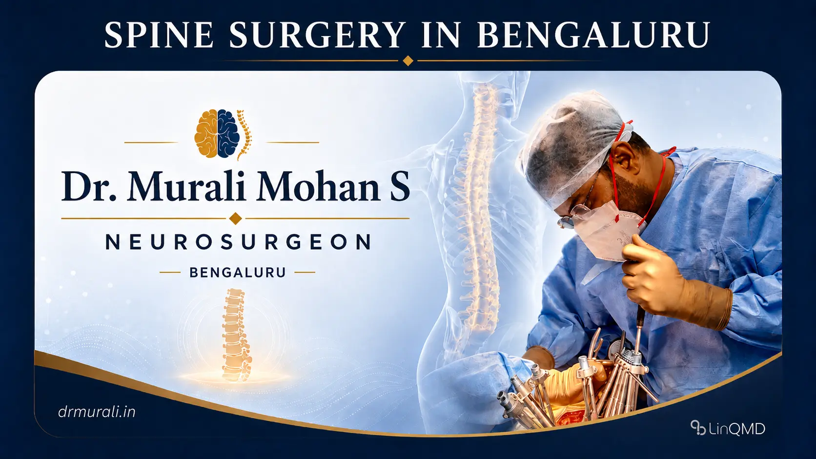
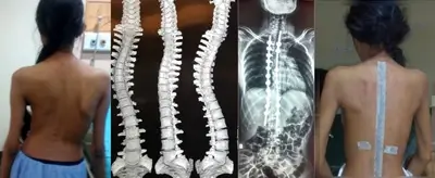
Dr. Murali Mohan provide spine surgery care in Bengaluru for cervical, lumbar, complex spine, and sp...
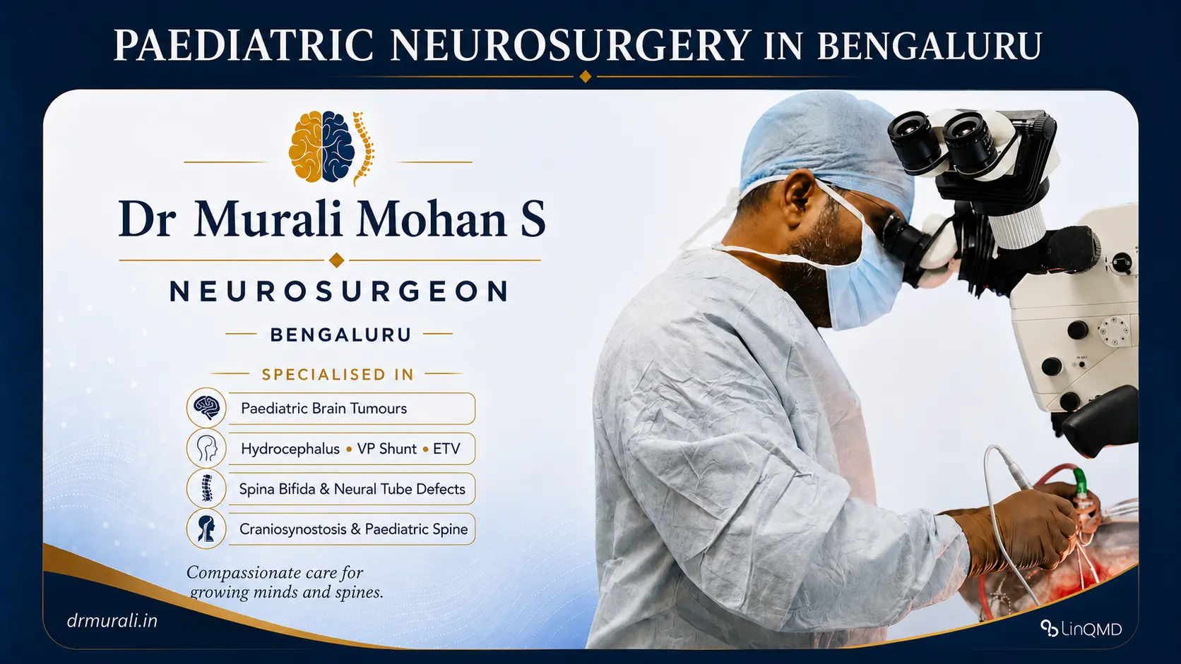
Dr. Murali Mohan S provides paediatric neurosurgical care in Bengaluru for brain, spine, and congeni...
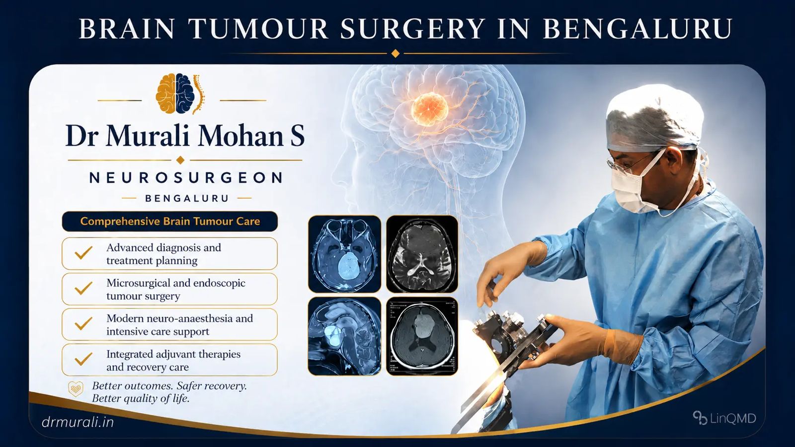
Brain tumour surgery is the treatment of abnormal growths within the brain or its coverings, carried...
Dr. Murali Mohan provide spine surgery care in Bengaluru for cervical, lumbar, complex spine, and sp...
Dr. Murali Mohan S provides paediatric neurosurgical care in Bengaluru for brain, spine, and congeni...
Brain tumour surgery is the treatment of abnormal growths within the brain or its coverings, carried...


Treatment of an AVM is focused on preventing bleeding and improving the patient’s quality of life. There are a few options, and sometimes they are used in combination:
Often, a combination is employed – for example, an AVM might be embolized first to reduce its size, then surgically removed; or partially embolized and then treated with radiosurgery. The approach depends on the AVM’s size, location, and the patient’s overall condition. Dr. Murali and the team will discuss the best plan in your case. The ultimate goal is to completely eliminate the AVM or at least block it off so that it cannot bleed with as low risk as possible.
Recovery varies, but most patients start feeling better the same day in the evening and are encouraged to walk. Those who don’t walk the same day are ensured that they walk the next day! With such quick recovery, the confidence builds up and they can get back to their routine life in a maximum of 2 weeks.
https://maps.app.goo.gl/CgH98dCkUPf1b3sA6
https://maps.app.goo.gl/yoQctYsXkDqb2j2FA