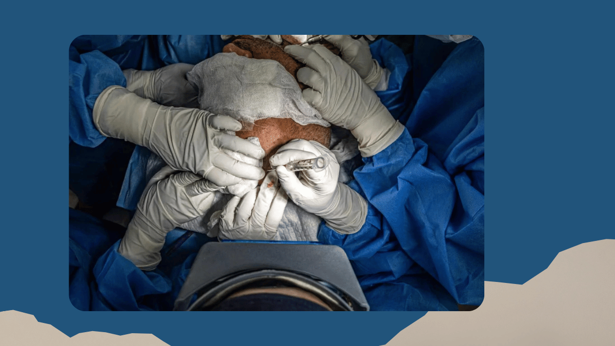
The skull base is the bony floor on which the brain sits, perforated by the arteries, veins, and cranial nerves that connect the brain to the face, the orbit, the inner ear, the pituitary gland, and the spinal cord. Every critical neurovascular structure that enters or exits the cranial cavity passes through, over, or near the skull base, which is why surgery in this region demands a level of anatomical knowledge, preoperative planning, and intraoperative precision that general brain surgery, even complex brain surgery, does not always require to the same degree. A meningioma on the surface of the brain convexity is managed very differently from the same histological tumour at the cavernous sinus or the clivus, where the tumour is surrounded by structures that control eye movement, facial sensation, swallowing, and the blood supply to the brainstem. My Fellowship training at Flinders Medical Centre, Adelaide, included specific exposure to skull base approaches and complex cranial surgery, and skull base surgery is one of the central areas of my practice at Gleneagles Fortis Hospital, Perumbakkam, Chennai.
Conditions Treated: What Arises at the Skull Base and Why Location Changes Everything
The conditions that bring patients to a skull base neurosurgeon are distinguished by their anatomical location as much as their pathological type, because the same histological tumour type at a different location in the skull base may require a completely different surgical approach, carry different risks, and produce different symptoms. The most common skull base conditions I manage at Gleneagles Fortis Hospital, Chennai include: pituitary tumours, which arise within or adjacent to the pituitary gland at the central skull base and typically present with hormonal disturbance, visual field loss from compression of the optic nerves, or headache; acoustic neuromas (vestibular schwannomas), which are benign tumours arising from the nerve sheath of the vestibulo-cochlear nerve at the internal auditory canal and cerebellopontine angle, presenting with progressive unilateral hearing loss, tinnitus, and sometimes balance disturbance; skull base meningiomas, which are meningiomas arising from the dural covering at the skull base in locations including the cavernous sinus, the tentorial notch, the clivus, the olfactory groove, and the sphenoid wing, each of which carries its own distinct surgical anatomy and risk profile; and chordomas, rare locally aggressive tumours arising from notochordal remnants at the clivus, the most challenging site in the skull base, where the tumour abuts the brainstem and basilar artery. The pillar guide on this profile describes skull base surgery as involving specialised approaches that access pathology while respecting critical structures. That sentence contains the essential truth of what makes this surgery demanding and what makes fellowship training and sustained operative experience the prerequisites for doing it well.
Surgical Planning, Approach Selection, and the Technology That Makes Skull Base Surgery Safer
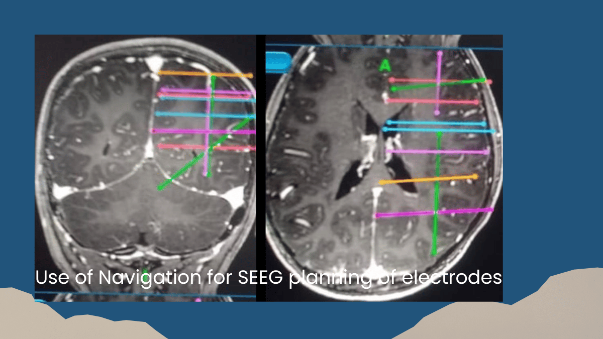
No skull base operation begins in the operating theatre. It begins with a detailed analysis of the imaging, the tumour's relationship to the surrounding neurovascular structures, and the surgical approach that will provide the safest and most complete access to the lesion while minimising manipulation of the structures that surround it. MRI with specific skull base sequences establishes the relationship of the tumour to the carotid arteries, the cavernous sinus, the cranial nerves, the brainstem, and the pituitary stalk. CT imaging shows the bony anatomy and the pneumatisation of the sinuses that influence approach selection. CT angiography maps the major arterial and venous structures where relevant. The surgical approaches used for skull base surgery each exploit specific anatomical corridors that provide access to different regions of the skull base with different trade-offs in terms of the structures that must be traversed or retracted to reach the target. For pituitary tumours, the endoscopic transsphenoidal approach, operating through the nostril and through the sphenoid sinus to reach the pituitary fossa, has become the standard for the majority of pituitary adenomas, avoiding any brain retraction entirely. For acoustic neuromas, the retrosigmoid approach through a small craniotomy behind the mastoid provides access to the cerebellopontine angle while preserving facial nerve function. Neuronavigation, which merges preoperative MRI and CT imaging with the patient's actual intraoperative position to allow real-time tracking of the surgeon's instruments in three-dimensional space, is used in virtually all skull base procedures at Gleneagles Fortis Hospital. Intraoperative neurophysiological monitoring of the facial nerve, the cranial nerves supplying eye movement, and brainstem auditory evoked potentials provides continuous real-time feedback that allows the surgical team to adjust technique when monitoring signals indicate proximity to critical structures. For patients in Chennai and across Tamil Nadu with skull base lesions, specialist neurosurgical assessment with the imaging review and surgical planning that complex skull base surgery requires is available at Gleneagles Fortis Hospital, Perumbakkam, without the need to travel to a tertiary centre outside the state.
To book a skull base surgery consultation with Dr. Nigel Peter Symss at Gleneagles Fortis Hospital, Perumbakkam, Chennai, contact the hospital directly. WhatsApp: +91 8861191019.
Written by Dr. Nigel Peter Symss, MBBS, DNB (Neurosurgery), Fellowship in Paediatric Neurosurgery (Jikei University School of Medicine, Tokyo, Japan, Oi-Sami Fellowship), Fellowship in Spinal Surgery, Functional Neurosurgery and Microneurosurgery (Flinders Medical Centre, Adelaide, Australia), HOD and Senior Consultant Neurosurgeon and Functional Neurosurgeon, Gleneagles Fortis Hospital, Perumbakkam, Chennai. 30+ years. 50,000+ patients.
Related reading
The Complete Guide to Brain and Spine Surgery: Everything Patients Need to Know
Brain Tumours: Symptoms, Diagnosis, and How Modern Surgery Has Changed Outcomes

Add a Comment