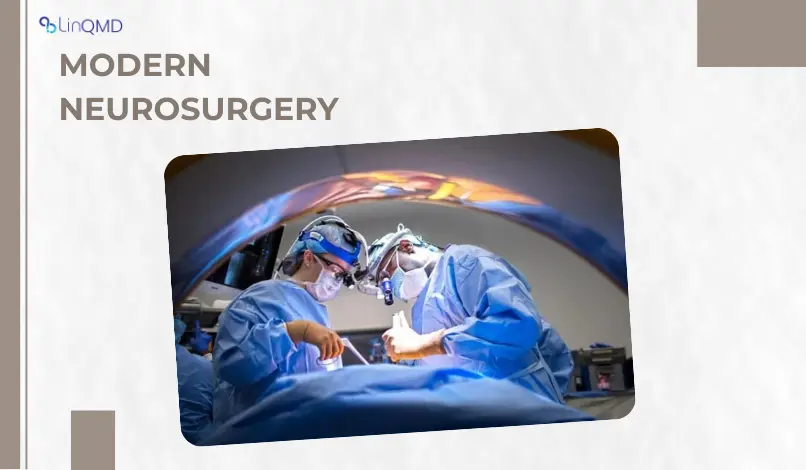
Brain tumours are among the most feared neurological conditions-yet today, with evolving diagnostics and surgical technology, the narrative is shifting. As someone who has spent decades working in neurosurgery, I want to share a deeper, patient-friendly look at what brain tumours are, how common they are, how outcomes are improving, and how my own work fits into this picture.
1. Incidence: How Common Are Brain Tumours?
Brain tumours are relatively rare, but their impact is significant. Globally, the age-standardized incidence rate of central nervous system (CNS) cancers is estimated at 3.5 per 100,000 people, with roughly 322,000 new cases each year.
According to GLOBOCAN 2022 data, around 248,000 annual deaths are attributed to brain and CNS cancers.
In India, primary brain tumour incidence is comparatively lower but increasing. A study from a tertiary hospital reported that brain and CNS tumours account for around 3.1% of all cancer incidences in their series. Age-adjusted rates in some population-based registries suggest a CNS tumour incidence of about 4.6 per 100,000 in parts of South Asia.
These numbers reflect both greater awareness and improved imaging access, especially in developing countries.
2. Types of Brain Tumours
Brain tumours are not a single disease-they vary widely in origin, behavior, and prognosis. Here are some of the major types:
Gliomas: These arise from glial cells. Subtypes include astrocytomas, oligodendrogliomas, and the most aggressive form: glioblastoma.
Meningiomas: Tumours that originate from the meninges (the membranes covering the brain) often benign but can cause serious problems due to location.
Pituitary tumours: Usually benign, these affect hormone regulation.
Other types: There are less common tumours, including ependymomas, medulloblastomas (more common in children), and metastatic lesions (tumours that have spread to the brain from other organs).
Each tumour type carries a different prognosis and treatment approach.
3. Outcomes Today: What Is the Prognosis?
Thanks to better technology, multidisciplinary teams, and more precise treatments, outcomes for brain tumour patients have improved-but they still vary widely.
For benign tumours (such as many meningiomas), patients often do very well after surgical removal. They may need regular imaging follow-up, but many can return to normal life with minimal neurological deficits. As I’ve seen in my own practice, rehabilitation and close monitoring are crucial.
Malignant tumours, especially high-grade gliomas like glioblastoma, remain challenging. Even with maximal treatment (surgery + radiotherapy + chemotherapy), median survival is often in the range of 12–15 months.
However, new tools help surgeons remove more tumour safely, leading to better “gross total resection” rates, which can translate into longer survival and better quality of life.
Long-term outcomes also depend on genetic and molecular features of the tumour. For example, molecular markers now guide therapy decisions in gliomas.
4. Technologies Today: What’s Changing in Brain Tumour Surgery?
The last decade has brought transformative technologies in brain tumour diagnosis and surgery, enhancing precision, safety, and outcomes. Here are some key advances:
1. Fluorescence-Guided Surgery (FGS)
Surgeons inject fluorescent dye (such as 5-ALA) that accumulates in tumour tissue. Under special light, cancerous tissue glows, helping us distinguish it from healthy brain.
Recent work with near-infrared (NIR) fluorescence shows that this can improve tumour margin detection, increasing gross total resection rates by 18–22%.
2. Intraoperative Navigation & Imaging
Much like a GPS for the brain, neuronavigation systems allow us to plan safe corridors to the tumour, minimizing damage to healthy areas.
Intraoperative MRI (iMRI) enables real-time scanning during surgery, so if any tumour remains, it can be removed in the same session.
3. Minimally Invasive Approaches
Endoscopic and endonasal surgery: For tumours near the skull base (e.g., pituitary), surgeons use an endoscope through the nasal route, avoiding large craniotomies and reducing recovery time.
Awake craniotomy: In certain cases (especially near “eloquent” brain areas), patients are kept awake, so we can test their function (speech, movement) while operating, reducing the risk of permanent neurological damage.
4. Optical & AI Technologies
Stimulated Raman Histology (SRH): This is a rapid, label-free optical imaging method that helps diagnose tumour tissue in real time during surgery.
Augmented Reality (AR) + Hyperspectral Imaging: Emerging systems like SLIMBRAIN overlay real-time imaging of tumour tissue onto the surgical field, helping surgeons visualize tumour boundaries with depth information.
AI-based intraoperative tools: For example, real-time tumour detection in intraoperative ultrasound using computer vision (e.g., YOLO11 architecture) is being developed to assist surgeons during operations.
These technological leaps are not just “nice to have”—they are making surgeries safer, faster, and more effective.
5. My Journey: Dr. Ravi Gopal Varma’s Connection to Brain Tumours
As a neurosurgeon with over 34 years of experience, I’ve had the privilege of walking alongside many patients in their brain tumour journeys. My training (MBBS, MS, MCh) and years in functional and stereotactic neurosurgery have shaped how I approach each case.
At Aster CMI Hospital, Bengaluru, where I currently serve as Lead Consultant Neurosurgery and Program Director, I help lead a Global Centre of Excellence in Neuroscience we have a multidisciplinary team of neuro-oncologists, neuro-imaging specialists, neuro-anesthetists, and neuro-rehab professionals.
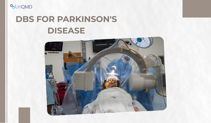
My philosophy is rooted in combining surgical precision with innovation. While my public profile is more often associated with movement disorders and DBS (Deep Brain Stimulation), I also perform brain tumour surgery, applying the latest technologies and guiding younger neurosurgeons in these complex procedures. My goal is to bring high-quality neurosurgical care to more people, improving survival and preserving function and dignity.
6. A Message for Patients and Families
If you or someone you love has been diagnosed with a brain tumour, here’s what you should know:
Get a second opinion: Given how varied brain tumours are, a specialised neuro-oncology centre can provide a more tailored evaluation.
Ask about advanced imaging and surgery: Techniques like fluorescence-guided surgery, intraoperative MRI, and endoscopic approaches can make a big difference.
Rehabilitation matters: Postoperative recovery isn’t just about removing the tumour. Physical therapy, neuro-rehab, and psychological support are essential.
Quality of life is a priority: Even when a tumour can’t be fully cured, reducing symptoms, preventing recurrence, and maintaining neurological function are extremely meaningful.
Final Thoughts
Brain tumours remain a challenging diagnosis but they are no longer an insurmountable one. Thanks to technological innovations and a multi-disciplinary approach, more patients are living longer, better lives than ever before.
In my work, every surgery is not just about removing a mass, it’s about restoring hope, functionality, and dignity. As a neurosurgeon, I feel deeply privileged to be part of that journey.
If you want to learn more or explore options, I’m here-not just as a surgeon, but as a guide who understands how deeply brain health intersects with life itself.
Dr. Ravi Gopal Varma is Lead Consultant Neurosurgery & Program Director, Global Centre of Excellence in Neurosciences, Aster Hospitals, Bangalore.

Add a Comment