India is the diabetes capital of the world, and the eyes pay a price that most people with diabetes do not fully appreciate until it is almost too late. Diabetic retinopathy is the most common cause of preventable blindness in working-age adults globally, and in a city like Bengaluru, where diabetes prevalence is rising steadily with urbanisation, sedentary work patterns, and changing diets, it is one of the conditions I manage most frequently at my clinic in Kasturi Nagar. I am Dr. Usha Shree, FVRS, Vitreo-Retina Specialist, and across 12 years and more than 10,000 patients, I have seen the full spectrum of this condition - from the earliest microaneurysms that are invisible to the patient and perfectly treatable, to the advanced proliferative disease that requires surgery and whose visual outcomes are far less predictable. The consistent finding is that diabetic retinopathy caught early is a very different disease from diabetic retinopathy caught late. This guide explains what every person with diabetes in Bengaluru needs to understand about their eyes.
How Does Diabetes Damage the Retina?
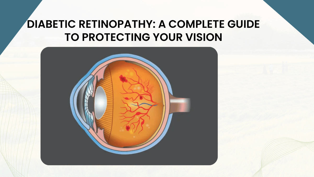
The retina is the light-sensitive tissue that lines the back of the eye - a thin, layered structure that converts the optical image formed by the lens into the electrical signals the brain interprets as vision. It is one of the most metabolically active tissues in the human body and one of the most densely vascularised for its size. This makes it exquisitely sensitive to the damage that sustained high blood sugar levels produce in small blood vessels throughout the body. The mechanism by which diabetes damages retinal blood vessels involves multiple interconnected pathways: glycation of vessel wall proteins, oxidative stress, inflammatory mediator release, and the accumulation of sorbitol within vessel wall cells through the polyol pathway. The cumulative effect is a progressive weakening of the walls of the tiny capillaries that supply the retina. The first visible consequence is the formation of microaneurysms - tiny balloon-like outpouchings of the capillary wall that leak fluid into the surrounding retinal tissue. As the disease progresses, capillaries close off entirely, producing areas of retinal ischaemia - tissue that is starved of oxygen and nutrients. The ischaemic retina then releases vascular endothelial growth factor, or VEGF, in an attempt to stimulate the growth of new blood vessels. But these new vessels - neovascularisation - are fragile, poorly constructed, and grow on the surface of the retina and into the vitreous gel filling the eye, where they bleed easily, contract, and can pull the retina away from the eye wall in a traction retinal detachment. Understanding this progression from vascular leak to ischaemia to neovascularisation is the framework through which every treatment decision in diabetic retinopathy is made.
The Four Stages of Diabetic Retinopathy
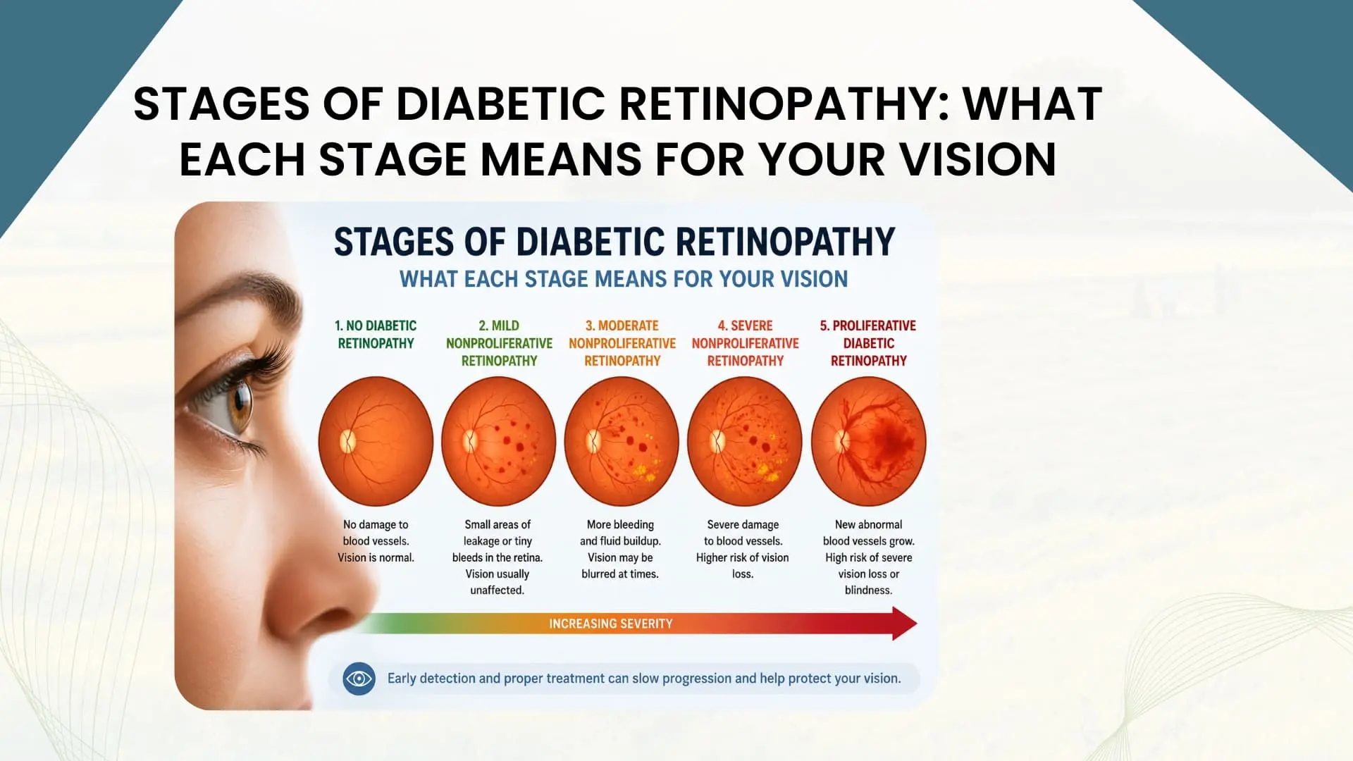
Diabetic retinopathy is classified into four stages that reflect the progression from early vascular changes to sight-threatening disease. Understanding these stages is clinically important because the treatment options and the urgency of intervention differ substantially between them.
Stage 1: Mild Non-Proliferative Diabetic Retinopathy (NPDR)
At this earliest stage, microaneurysms are the only finding on fundus examination. The patient typically has no visual symptoms whatsoever. This is the stage where intervention in the underlying diabetes - achieving tighter blood sugar control, managing blood pressure, and addressing lifestyle factors - has the greatest potential to slow or halt progression. The absence of symptoms is not reassurance that nothing is wrong; it is the reason that annual retinal screening for every person with diabetes is a non-negotiable clinical recommendation.
Stage 2: Moderate Non-Proliferative Diabetic Retinopathy
Microaneurysms are joined by retinal haemorrhages, hard exudates from leaking protein, and cotton wool spots from areas of focal ischaemia. Vision may still be normal if the macula - the central area of the retina responsible for reading and detailed vision - is not involved. However, the risk of progression is meaningfully higher at this stage, and closer monitoring intervals are required.
Stage 3: Severe Non-Proliferative Diabetic Retinopathy
This stage is defined by widespread retinal haemorrhages in all four quadrants of the retina, venous beading - where the retinal veins develop an irregular, bead-like appearance from their engorgement - and intraretinal microvascular abnormalities. It is a stage of significant retinal ischaemia. The risk of progressing to proliferative disease within one year is substantial, and treatment planning typically begins at this stage even in the absence of new vessel formation.
Stage 4: Proliferative Diabetic Retinopathy (PDR)
New blood vessels have grown on the surface of the retina or optic disc. This is the stage at which the greatest risk of severe visual loss exists. The fragile new vessels bleed into the vitreous - causing sudden floaters, a dark veil over vision, or complete loss of vision. Fibrovascular membranes attached to these vessels can contract and detach the retina from the eye wall, a traction retinal detachment that requires surgical repair. Proliferative diabetic retinopathy requires urgent specialist treatment - it is not a condition that can be safely observed.
Diabetic Macular Oedema: When the Centre of Vision is Affected
Diabetic macular oedema, or DME, is the accumulation of fluid in the macula - the central region of the retina that is responsible for reading, recognising faces, and performing all detailed visual tasks. DME can occur at any stage of diabetic retinopathy, including in the early stages when the peripheral retina shows only minimal changes. It is the most common cause of vision loss in people with diabetes, and it is often the first manifestation of diabetic retinal disease that the patient notices, presenting as blurring of central vision or difficulty reading. The driving mechanism is the breakdown of the blood-retinal barrier from leaking microaneurysms and diseased capillaries, allowing fluid and lipoproteins to accumulate in the layers of the macular tissue. On optical coherence tomography (OCT) - the imaging technology that has transformed the diagnosis and monitoring of DME - the fluid appears as dark cystic spaces within the retinal layers, and its distribution and extent guide both the severity classification and the treatment approach. Diabetic macular oedema is now the most common indication for anti-VEGF injections in retinal practice, and the visual outcomes for patients who receive treatment promptly are substantially better than they were before this treatment class became available.
Symptoms - What Diabetic Retinopathy Feels Like
One of the most clinically important facts about diabetic retinopathy is that early stages produce no symptoms at all. The mild and moderate non-proliferative stages, which are the stages most amenable to intervention and prevention of progression, are invisible to the patient. There is no pain, no visual change, and no warning signal from the body that anything is developing in the eye. This is why the annual fundus examination is not optional for people with diabetes - it is the only way to detect a condition that will announce itself with symptoms only when it has already reached a more advanced and harder-to-treat stage.
The symptoms that patients do eventually report, when disease has progressed, include: blurred vision that is often fluctuating with blood sugar levels; floaters - the sudden appearance of spots, threads, or a shower of dark dots in the vision, which may represent a vitreous haemorrhage from a bleeding new vessel; a dark curtain or shadow over part of the visual field, which may represent a retinal haemorrhage or detachment; distortion of straight lines, particularly when macular oedema has developed; and difficulty with colour discrimination in more advanced disease. Any of these symptoms in a person with diabetes warrants same-day or next-day retinal specialist review, not watchful waiting.
Risk Factors - Who Is Most at Risk?
The two most powerful determinants of diabetic retinopathy risk are the duration of diabetes and the degree of glycaemic control. Someone who has had poorly controlled type 2 diabetes for fifteen years faces a substantially higher retinopathy risk than someone who has had well-controlled type 1 diabetes for five years. This means that for many people newly diagnosed with type 2 diabetes in Bengaluru - where the average age at diagnosis has been falling - the metabolic damage that precedes the formal diagnosis may already have been accumulating for years before the diabetes was identified.
The additional risk factors that compound the probability of developing or worsening diabetic retinopathy include: hypertension, which independently damages retinal vessels and amplifies the effect of hyperglycaemia; dyslipidaemia, where elevated serum lipids are associated with hard exudate formation in the macula; nephropathy, where the presence of diabetic kidney disease is a strong indicator that microvascular disease is systemic and likely affecting the retina; pregnancy in a diabetic woman, where rapid fluctuations in metabolic control can accelerate retinopathy progression significantly; anaemia, which worsens retinal ischaemia; smoking; and obesity. The patient with diabetes, hypertension, elevated cholesterol, and a long duration of poorly controlled blood sugar is the patient I see most commonly presenting with significant retinopathy in my Bengaluru practice.
How Diabetic Retinopathy Is Diagnosed
Accurate diagnosis and staging of diabetic retinopathy requires a systematic examination of the retina by a specialist with the imaging technology to see what the clinical examination alone may not reveal.
Dilated Fundus Examination
The clinical fundus examination, performed after pupil dilation with eye drops, is the foundational examination in diabetic retinopathy evaluation. Using an indirect ophthalmoscope and a slit lamp with a condensing lens, I examine the retina systematically - from the optic disc and major arcades through the peripheral retina - identifying the haemorrhages, exudates, new vessels, and other findings that determine the stage of the disease. In my practice, no diabetic retinopathy assessment is complete without full fundus examination through a dilated pupil.
Optical Coherence Tomography (OCT)
OCT is the imaging modality that has most transformed the management of diabetic macular oedema. It produces cross-sectional images of the retinal layers with micrometre-level resolution, allowing me to identify the location, extent, and type of macular fluid accumulation with precision that was not previously achievable. OCT also monitors the response to treatment: after each anti-VEGF injection, the OCT scan at the next visit shows whether the fluid has resolved, partially resolved, or recurred, and this guides the decision about continuing, modifying, or pausing treatment.
Fundus Fluorescein Angiography (FFA)
FFA involves the injection of a fluorescent dye into a vein in the arm and then photographing the retinal vasculature as the dye passes through. This reveals the pattern of vascular leakage, identifies areas of capillary non-perfusion - the ischaemic zones that drive neovascularisation - and precisely maps the distribution of new vessel formation. FFA is particularly valuable for treatment planning before laser photocoagulation and for identifying the extent of ischaemia that determines whether panretinal photocoagulation is indicated.
OCT Angiography (OCT-A)
OCT-A is a newer non-invasive imaging modality that maps retinal vasculature without the injection of dye. It provides exquisite detail of the superficial and deep retinal capillary plexuses and the foveal avascular zone, identifying capillary dropout and microaneurysms with resolution that FFA cannot achieve. It is increasingly used in my practice for early detection and for monitoring subtle vascular changes that precede clinically apparent disease.
Treatment of Diabetic Retinopathy
The treatment approach in diabetic retinopathy is determined by the stage of the disease and whether diabetic macular oedema is present. In all cases, optimisation of the systemic control of diabetes, hypertension, and dyslipidaemia is the most important long-term protective intervention available - no retinal treatment can fully compensate for continued vascular damage from uncontrolled metabolic disease.
Anti-VEGF Injections
Anti-VEGF medications - including ranibizumab, bevacizumab, and aflibercept - are injected into the vitreous cavity of the eye through the sclera in a minor outpatient procedure performed under topical anaesthesia. They work by neutralising vascular endothelial growth factor, the molecule responsible for both the vascular leakage driving macular oedema and the neovascularisation driving proliferative disease. Anti-VEGF injections have become the first-line treatment for centre-involving diabetic macular oedema, replacing laser therapy as the primary modality because of their superior outcomes for central visual acuity. Anti-VEGF injections for diabetic retinopathy require a structured treatment protocol - typically monthly injections for the initial loading phase, followed by a monitoring-and-treat or treat-and-extend schedule - and the visual results in patients who receive prompt, consistent treatment are substantially better than those treated late or inconsistently.
Laser Photocoagulation
Laser treatment remains an important component of diabetic retinopathy management. Focal laser is applied to specific leaking microaneurysms outside the central macula, targeting the sites of fluid leakage and reducing the total macular oedema burden - often used in combination with anti-VEGF for clinically significant macular oedema. Panretinal photocoagulation (PRP) involves applying laser burns across the peripheral retina to destroy the ischaemic tissue that is producing VEGF, thereby reducing the stimulus for neovascularisation. PRP remains the primary treatment for proliferative diabetic retinopathy in many settings, though it is increasingly combined with anti-VEGF to accelerate the regression of new vessels while the peripheral laser burns consolidate.
Vitrectomy Surgery
When vitreous haemorrhage does not clear spontaneously, when traction retinal detachment has developed or is threatening the macula, or when dense fibrovascular membranes require removal, vitrectomy surgery becomes necessary. This is a microsurgical procedure in which the vitreous gel and associated fibrovascular membranes are removed from the eye using fine instruments introduced through small incisions in the sclera. In my practice at Kasturi Nagar, Bengaluru, with more than 400 surgical cases performed, vitrectomy for advanced diabetic complications is one of the most complex and demanding procedures in retinal surgery - but one that, when performed at the appropriate stage, can preserve or restore useful vision in patients who would otherwise face profound visual loss. Early specialist referral before traction retinal detachment has involved the macula is the single most important factor in determining the visual outcome of vitrectomy for diabetic complications.
Prevention and Protecting Your Vision With Diabetes
Prevention of diabetic retinopathy begins with the management of diabetes itself, and the evidence for this is unambiguous: the DCCT and UKPDS trials demonstrated decades ago that intensive glycaemic control reduces the incidence and progression of diabetic retinopathy by 50 to 70 percent compared with conventional control. In practice, this means targeting an HbA1c of 7 percent or below where this can be achieved safely and without excessive hypoglycaemia, controlling blood pressure to below 130/80 mmHg, and treating dyslipidaemia. Beyond metabolic control, regular physical activity, maintenance of a healthy body weight, smoking cessation, and a diet that is low in refined carbohydrates and rich in antioxidant nutrients are all protective.
The other pillar of prevention is systematic screening. Every person with type 1 diabetes should have a dilated fundus examination within the first five years of diagnosis and annually thereafter. Every person with type 2 diabetes should have a dilated fundus examination at the time of diagnosis and annually thereafter - because many people with type 2 diabetes have had the condition for years before it is diagnosed, and may already have retinopathy at the time of their first formal blood sugar test. For patients with established retinopathy, the screening interval is shortened to reflect the stage of disease. Regular retinal examination is the only intervention that finds diabetic retinopathy before it steals your vision.
When to See a Retina Specialist in Bengaluru
If you have diabetes, the threshold for seeking a retinal review should be low. Specific situations that warrant prompt specialist assessment include: any new visual symptom - blurring, floaters, a dark shadow or curtain, or distorted lines; a diagnosis of diabetes in a patient who has never had a retinal examination; a patient whose last retinal examination was more than twelve months ago; pregnancy in a diabetic woman, who should be seen before conception or in the first trimester and monitored closely throughout; and any patient whose systemic physician or endocrinologist has found significant HbA1c levels, new hypertension, or signs of diabetic nephropathy.
In my practice in Kasturi Nagar, Bengaluru, I see patients across the full spectrum of diabetic eye disease and have the imaging technology - OCT, FFA, wide-field fundus photography - and the procedural capability - anti-VEGF injections, laser photocoagulation, and vitrectomy surgery - to provide the complete range of management that diabetic retinopathy requires. The most common regret I hear from patients is that they wished they had come sooner. The most common finding that I can act on most effectively is the one I see before the patient has any symptoms at all.
To book a consultation with Dr. Usha Shree at Kasturi Nagar, Bengaluru, call +91 9663358193.
Written by Dr. Usha Shree, MBBS (RGUHS, 2007), DNB Ophthalmology (NBE, 2012), FICO (ICO, 2015), FVRS (RGUHS, 2016), Vitreo-Retina Specialist, Kasturi Nagar, Bengaluru. 12 years, 10,000+ patients. Phone: +91 96633 58193.
Related reading
Diabetic Retinopathy: Early Symptoms, Risk Factors, and Treatment Options
Stages of Diabetic Retinopathy: What Each Stage Means for Your Vision

Add a Comment